Abstract: Objective: To evaluate how surfactant-mediated biofilm disruption by hops beta (β)-acids extract influences stannous fluoride (SnF2) penetration and interaction with extracellular metabolites, including lipopolysaccharide (LPS). Methods: In two studies, oral biofilms were formed in vivo on artificial enamel surfaces mounted in dental appliances worn by volunteers and subsequently treated ex vivo under controlled conditions. Across the studies, biofilms were treated with supernatants of various commercial 0.454% SnF2 dentifrices containing: (1) pyrophosphate; (2) potassium nitrate (KNO3) and pyrophosphate; (3) tripolyphosphate; and (4) gluconate/citrate–stabilized 0.454% SnF2 (Crest) with and without hops β-acids extract (Activ Bio-Cleanser™). Both studies used phosphate-buffered saline as a negative control (NC). Confocal laser scanning microscopy with component-specific fluorescent probes was used to quantify Sn and LPS distribution relative to bacterial biomass, biofilm thickness, and extracellular polymeric substance (EPS) structure. Image analysis was used to quantify Sn and LPS signal within bacterial regions, normalized to bacterial volume or area; statistical comparisons were made across conditions. Results: SnF2 uptake and penetration varied significantly across formulations. In study 1, the rank order from greatest to least for Sn deposition and LPS neutralization was: gluconate/citrate > KNO3 + pyro > pyro > NC (P < 0.05 between groups). In study 2, the rank order from greatest to least was: gluconate/citrate + hops > gluconate/citrate > tripolyphosphate and KNO3 + pyro > NC (P < 0.05 between groups). Some stabilized SnF2 systems exhibited limited penetration, consistent with diffusion barriers imposed by dense EPS matrices or over-stabilization reducing bioavailability. Gluconate/citrate–stabilized SnF2-only treated biofilms showed higher deposition per unit bacteria than other SnF2 technologies but uniformly exhibited the majority of uptake in surface regions. In the formulation containing both hops and SnF2, increased Sn penetration and reduced detectable biofilm-associated LPS was observed, consistent with enhanced access of SnF2 to extracellular plaque components following surfactant-enabled biofilm modification. In this case, Sn penetration achieved high relative deposition per unit bacteria in the treated regions throughout the biofilm. Conclusions: Hops β-acids extract acts as a co-surfactant in toothpaste, capable of modifying EPS structure to improve accessibility and interaction of SnF2 with extracellular metabolites within oral biofilms. The observed changes in Sn penetration and biofilm-associated, extracellular LPS are consistent with enhanced cleaning with reduced concentration of known tissue irritants. Clinical Implications: These findings provide a mechanistic rationale for formulation strategies that enhance SnF2 accessibility within adherent oral biofilms through surfactant-based modification of EPS supporting more efficient biofilm management during oral hygiene and providing a basis for selecting toothpastes based on their efficacy.
Keywords: surfactant, extracellular polymeric substances, hops β-acids, dental hygiene, toothpaste
Stannous fluoride (SnF2) is widely formulated into toothpastes and has a long history of use in oral care. Originally utilized in the 1950s for the prevention of dental decay,1,2 SnF2 in contemporary formulations offers additional benefits, such as treatment of plaque-associated gingivitis and reduction of dentin hypersensitivity.3-5 Antiplaque/antigingivitis efficacy is derived from antibacterial actions of SnF2, including detoxification of the extracellular endotoxins in microbial biofilms.6-9 As with other toothpaste ingredients, the effectiveness of SnF2 depends on penetration and retention into target substrates, including adherent oral biofilms.
A distinct challenge for SnF2 toothpaste design is the need to balance chemical stability with bioavailability.10 The stannous (Sn2+) portion of SnF2 is prone to oxidation into ineffective stannic, and SnF2 must be protected from destabilization reactions during formulation and storage. However, the stabilization approaches used to protect Sn2+ can influence diffusion, reactivity, and retention (collectively bioavailability) in the oral cavity, creating an inherent formulation trade-off.11 Successful formulation of SnF2 thus requires the balancing of stability with bioavailability.
This trade-off is particularly relevant in adherent oral biofilms in which bacteria reside within a protective extracellular polymeric substance (EPS) matrix that promotes cohesion, adhesion, and mechanical tenacity on tooth surfaces that can further accumulate tissue-irritating metabolites, including lipopolysaccharides (LPS) and volatile sulfur compounds.12 As with virtually all microbial populations in nature, oral microbes reside in the oral cavity as 3-dimensional coatings on surfaces, described as biofilms.13,14 To form biofilms, bacteria produce matrix components that act to “glue” the films together and ensure their tenacity to surfaces.15 Biofilm matrices are significant contributors to survival of bacterial species16-20 by presenting a limitation to effective physical removal during hygiene. The EPS-rich matrix can also function as a diffusion barrier that limits the penetration of agents into deeper regions of the biofilm.15,21,22 As a result, agents may show preferential accumulation near the biofilm surface, with limited penetration into the internal biofilm architecture.23 In the context of SnF2, these biofilm transport constraints may interact with formulation stabilization choices to determine how much stannous reaches, penetrates, and is retained within the adherent oral biofilms—namely SnF2 bioavailability.
Within the broader framework of this special issue, this article addresses the hypothesis that formulation-enabled modification of EPS structure can reduce diffusion barriers and improve delivery of subsequently applied active ingredients during toothbrushing. Prior research has shown that hops beta (β)-acids extract, when delivered as part of an oral care formulation strategy, can solubilize and disperse EPS in ex vivo–treated oral biofilms, consistent with a surfactant-based, physical–chemical cleansing mechanism.24 In this context, hops are positioned as surfactants that loosen EPS-rich adherent oral biofilms, thereby enabling improved access for active ingredients such as SnF2.
A second gap addressed by this article is the need for penetration metrics that are robust to differences in biofilm thickness and biomass. To enable depth-resolved assessment of delivery within complex biofilm matrices, Procter & Gamble laboratories have successfully developed and applied specific fluorescent staining procedures for confocal laser scanning microscopy (CLSM) to help visualize effects of formulations on in situ–derived biofilms.25 This includes a modified rhodamine B derivative, which is a novel stain developed and patented to provide specific visualization of stannous in complex matrices with excitation at 520 nm to 580 nm and analysis at 560 nm to 660 nm.25 These imaging tools support quantitative evaluation of stannous distribution relative to bacterial biomass and biofilm structure, enabling comparisons of “delivery efficiency” across biofilm states and formulation types.
Accordingly, the purpose of this study was twofold. First, uniquely stabilized commercial SnF2 toothpastes were compared for stannous uptake, penetration, and retention within in situ–generated adherent oral biofilms to assess the influence of formulation differences. Second, an evaluation assessed whether incorporation of a hops β-acids extract into a gluconate/citrate–stabilized, stannous-containing toothpaste could enhance subsequent stannous uptake and penetration into adherent oral biofilms under controlled ex vivo conditions. In addition, the study design supported assessment of formulation efficacy for removal of extracellular biofilm–associated components related to tissue irritation, LPS, using in situ–derived biofilms.
Methods and Materials
Overview of Experimental Approach
This study used in situ–generated adherent oral biofilms formed on hydroxyapatite (HA) discs mounted on removable oral splints and then treated ex vivo to enable assessment of Sn uptake, penetration, and retention from toothpaste supernatants. Biofilms were: (1) exposed to supernatants prepared from toothpastes to compare delivery behavior under different formulation conditions (Sn stabilization and hops β-acids extract content), and (2) assessed using CLSM with component-specific fluorescent probes to quantify Sn and LPS distribution relative to EPS distribution and bacterial biomass.
Biofilm Donors and Participant Characteristics
In situ biofilms were collected from a panel meeting the following criteria: between 18 to 50 years of age, good general health, free of active oral disease, minimum of 18 natural teeth, and demonstrated measurable plaque accumulation (defined as a score of 2–3 on the Turesky Modified Quigley Hein Plaque Index).26,27 The protocol was approved by the Procter & Gamble site safety group, and the study followed Good Clinical Practices. All participants provided written informed consent prior to participation.
In Situ Biofilm Generation and Substrate Preparation
Figure 1 shows the in situ plaque biofilm model, which was established based on minimal modification to the bovine dentin groove models.23,26,28 Instead of bovine, HA discs (Bei’erkang) were used to more closely approximate human enamel mineral composition and provide disc-to-disc uniformity and the necessary structural integrity for groove machining. Discs were 6 mm in diameter and 1 mm in thickness, cut with three parallel grooves perpendicular to the disc surface (the middle groove being 500 µm in both depth and width, and the bilateral grooves being 300 µm in both depth and width). HA discs were fixed onto a customized removeable oral splint on buccal surfaces of molars and premolars for defined periods, allowing for natural biofilm accumulation for 48 hours under normal conditions.
Participants were instructed to wear the oral splint except during meals (during which time the splint would be stored in an opaque container in humid conditions without soaking in liquid) and when performing oral hygiene procedures. During the plaque accumulation phase, participants brushed their teeth using a provided commercial 0.243% sodium fluoride toothpaste (Crest® Cavity Protection, The Procter & Gamble Company, crest.com) and a manual toothbrush (Crest® Velvet Soft Bristle, The Procter & Gamble Company) twice daily during the study. Participants were instructed not to use any other non-study oral hygiene products (eg, dental floss, oral rinse) over the course of the study.
Test Materials and Treatment Conditions
Biofilms were treated with supernatants of commercial 0.454% SnF2 toothpastes from three manufacturers along with a formulation incorporating hops β-acids extract. The hops β-acids extract was supplied as a commercially available botanical extract (Beta Bio 45%, Hopsteiner, hopsteiner.us), containing approximately 45% β-acids by weight. Phosphate-buffered saline (PBS) was used as a negative control in both studies. The treatments in each study were as follows:
Study 1
a. Negative control: PBS (1x, sterile-filtered, ThermoFisher Scientific Inc., thermofisher.com)
b. SnF2-pyrophosphate: 0.454% SnF2 toothpaste (Colgate Total Whitening, Colgate-Palmolive, colgate.com) supernatant
c. SnF2-pyrophosphate: 0.454% SnF2 toothpaste (Colgate Total Clean Mint, Colgate-Palmolive) supernatant
d. SnF2-potassium nitrate (KNO3)/pyrophosphate: 0.454% SnF2 toothpaste (Colgate Total Active Penetration Whitening, Colgate-Palmolive) supernatant
e. SnF2-KNO3/pyrophosphate: 0.454% SnF2 toothpaste (Colgate Total Active Penetration Clean Mint, Colgate-Palmolive) supernatant
f. SnF2-gluconate/citrate: 0.454% SnF2 toothpaste (Crest Pro-Health Clean Mint, The Procter & Gamble Company) supernatant
g. SnF2-gluconate/lactate/hexametaphosphate: 0.454% SnF2 toothpaste (Crest Pro-Health Advanced Deep Clean, The Procter & Gamble Company) supernatant
Study 2
A. Negative control: PBS (1x, sterile-filtered, ThermoFisher Scientific Inc.)
B. SnF2-KNO3/pyrophosphate: 0.454% SnF2 toothpaste (Colgate Total Gum Protect Whitening, Colgate-Palmolive) supernatant
C. SnF2-tripolyphosphate: 0.454% SnF2 toothpaste (Parodontax Complete Protection, Haleon, parodontax.com) supernatant
D. SnF2-gluconate/citrate: 0.454% SnF2 toothpaste (Crest Pro-Health Gum Detoxify Deep Clean, The Procter & Gamble Company) supernatant
E. SnF2-gluconate/citrate: 0.454% SnF2 toothpaste (Crest Pro-Health Advanced Bacteria Protection, The Procter & Gamble Company) supernatant
F. SnF2-gluconate/citrate-hops: 0.454% SnF2 toothpaste with 0.15% hops β-acids (Crest Gum Detoxify with Activ Bio-Cleanser™, The Procter & Gamble Company) supernatant
Supernatants were prepared prior to use by mixing 5 g of treatment/toothpaste with 15 mL of ultra-pure water using a homogenizer, followed by centrifugation at 10,000 rpm for 20 minutes. PBS was used as supplied.
Biofilm Treatment and Dispersion Protocols
Following the 48-hour wear period, the biofilm-coated HA discs were removed from the splint and gently rinsed with PBS solution. In situ–derived biofilms on HA discs were treated ex vivo by quiescent immersion in 500 μL of the assigned test solution/toothpaste slurries for 2 minutes. Treated and rinsed HA discs were stored at 4°C prior to microscopy analysis.
Confocal Laser Scanning Microscopy and Fluorescent Labeling
Biofilm structure and composition were evaluated using CLSM. Before imaging, treated biofilms were stained with fluorescent probes selected to enable separate visualization of key biofilm components, as noted in Table 1. Multichannel CLSM imaging was performed to capture spatial distribution and co-localization of bacterial cells, EPS matrix, and mineral components within the biofilm. Six selected fields were considered as representative of the whole sample after the experienced observer’s general examination.26 Optical sectioning enabled assessment of biofilm thickness and internal structure.
All samples were analyzed in 35-mm glass-bottom dishes (Mattek, mattek.com) by CLSM (TCS SP8 AOBS, Leica Microsystems, leica-microsystems.com) using a 40x dry objective (HCX PL APO/bd.BL 40.0, Leica Microsystems). The confocal system consisted of a Leica DM6000B upright microscope and Leica DMIRE2 inverted microscope. Leica™ confocal software LAS AF3.3.0 provided an interface for multidimensional image series acquisitions, processing, and analysis, which included 3D image reconstruction and measurement, physiological recording and analysis, time-lapse, fluorochrome co-localization, spectral inmixing, and multicolor restoration. CLSM image stacks were analyzed to assess changes in biofilm coverage, thickness, and compositional distribution following treatment. Qualitative comparisons focused on EPS integrity, biofilm cohesion, and adhesion to enamel surfaces, while quantitative measurements were used where appropriate to support comparison across treatments.
Fluorescence Probe Staining Microscopy Parameters
Application of fluorescent probes for histomorphological CLSM analysis has been described previously.23,26,28 An “ion fluorescent probe” refers to a fluorescent probe that specifically binds to one kind of ion/molecule and emits fluorescence at a certain wavelength. HA/biofilm specimens were soaked in 100 µl respective fluorescence solution and incubated at room temperature in the dark for 15 minutes. Table 1 shows probes used in this study.
For the primary penetration analyses, specimens were stained to evaluate bacterial content and Sn2+ signal, using SYTO-9 (bacterial label) and a modified rhodamine B derivative for Sn visualization.25 The analysis approach normalized stannous signal to bacterial content to support comparative interpretation across specimens.
Image Sampling Strategy and Thickness Measurement
For each specimen, three selected fields per sample were considered as representative of the whole sample after the observer’s general examination. The biofilm volume was determined from the confocal measurement. This volume was divided by the area of the scan to determine the mean thickness of the biofilm for each specimen.
Statistical Analysis
Each treatment group was evaluated with multiple fields of view on multiple specimens, and treatment differences were summarized using Student-Newman-Keuls multiple-comparison groupings (treatments in different statistical groups differ at P < 0.05). An average value for each HA disc was made based on three fields per disc. Then the average treatment result was obtained by averaging the results from the three treated discs together. Analyses were, therefore, done at the HA disc level (n = 3).
Results
Two ex vivo studies evaluated formulation-dependent differences in Sn deposition and LPS neutralization in in situ–derived adherent oral biofilms treated under standardized conditions using fluorescent markers and CLSM. Sn deposition for studies 1 and 2 is illustrated in Figures 2a and 2b, respectively, while Sn penetration for studies 1 and 2 is depicted in Figures 3a and 3b, respectively. Finally, LPS neutralization for studies 1 and 2 is demonstrated in Figures 4a and 4b, respectively.
In study 1, bacteria-normalized Sn deposition differed across the seven treatment conditions, as reported in Table 2, spanning 0.03 for the negative control (treatment a) to 1.32 for SnF2-gluconate/citrate Crest Pro-Health Clean Mint (treatment f). The SnF2-pyrophosphate treatments were the lowest tier of efficacy (0.38 and 0.46, Colgate Total Whitening and Clean Mint, treatments b and c, respectively). The next tier was formed by the SnF2-KNO3/pyrophosphate treatments (0.89 and 0.72, Colgate Total Active Penetration Whitening and Clean Mint, treatments d and e, respectively), where there was improvement versus pyrophosphate-only Sn stabilization. The highest tier of efficacy in study 1 consisted of the SnF2-gluconate/citrate toothpaste and the SnF2-gluconate/lactate/hexametaphosphate toothpaste (1.32 and 1.28, Crest Pro-Health Clean Mint and Crest Pro-Health Advanced Deep Clean Mint, treatments f and g, respectively). Products within each tier were not significantly different, whereas products in different tiers were significantly different (P < 0.05).
The bacteria-normalized LPS neutralization results in study 1 exhibited a parallel tiering, ranging from 0.18 for the negative control (treatment a) to 1.46 for Crest Pro-Health Clean Mint (treatment f). Treatments b and c again formed the lowest tier (0.69 and 0.77, respectively), and treatments d and e formed the next tier (1.17 and 1.04, respectively). The highest tier again comprised treatments f and g (1.46 and 1.35, respectively). Again, products within tiers were not significantly different, while products in different tiers were significantly different (P < 0.05).
In study 2, bacteria-normalized Sn deposition differed across the six treatment conditions (Table 3), spanning 0.06 for the negative control (treatment A) to 2.18 for SnF2-gluconate/citrate-hops (Crest Gum Detoxify with Activ Bio-Cleanser, treatment F). The SnF2-KNO3/pyrophosphate and SnF2-tripolyphosphate treatments (1.25 and 1.34, Colgate Total Gum Protect Whitening and Parodontax Complete Protection, treatments B and C, respectively) were the lowest tier of efficacy. The next tier was formed by the SnF2-gluconate/citrate treatments (1.69 and 1.82, Crest Pro-Health Gum Detoxify Deep Clean and Crest Pro-Health Advanced Bacteria Protection, treatments D and E, respectively), where there was improvement versus KNO3/pyrophosphate or tripolyphosphate Sn stabilization. The highest tier of efficacy in study 2 consisted of the SnF2-gluconate/citrate-hops toothpaste (2.18, Crest Gum Detoxify with Activ Bio-Cleanser, treatment F). Products within each tier were not significantly different. Products in different tiers were significantly different (P < 0.05).
The bacteria-normalized LPS neutralization results in study 2 also exhibited a parallel tiering, ranging from 0.34 for the negative control (treatment A) to 1.69 for Crest Gum Detoxify with Activ Bio-Cleanser (treatment F). Treatments B and C formed the lowest tier (0.73 and 0.76, respectively), and treatments D and E formed the next tier (0.92 and 0.98, respectively). The highest tier comprised treatment F (1.69). Again, products within tiers were not significantly different, while products in different tiers were significantly different (P < 0.05).
Across both studies, the lowest bacteria-normalized values for both endpoints occurred in the respective negative control groups (treatment a in study 1; treatment A in study 2), while the highest tiers were observed among the top-performing treatments in each study (treatments f/g in study 1; treatment F in study 2). Results for the hops-containing, SnF2 toothpaste stabilized with gluconate/citrate (treatment F, study 2) were universally the highest observed across both studies. In both studies, the statistical tiering for neutralized LPS closely tracked the tiering observed for bacteria-normalized Sn deposition, with intermediate comparator groups clustering together and the upper-tier formulations clustering. Taken together, the two studies demonstrate that dentifrice formulations containing the same level of 0.454% SnF2 can differ substantially in bacteria-normalized Sn deposition into adherent oral biofilms and in the associated bacteria-normalized neutralized LPS results under controlled ex vivo conditions.
Discussion
This research used CLSM histomorphological analysis to evaluate formulation-dependent differences in Sn deposition and penetration within in situ–derived adherent oral biofilms treated ex vivo and to quantify associated neutralization of biofilm-associated, extracellular LPS. Across two studies, Sn deposition and LPS neutralization per unit bacteria showed consistent tiering patterns across treatment sets, demonstrating that dentifrice formulations can differ substantially in delivery and interaction behavior in adherent oral biofilms under controlled conditions despite the formulations being comprised of similar levels of SnF2 (0.454%). In study 1, Crest Pro-Health formulations formed the highest tier relative to the other variants, while in study 2 the hops-containing, gluconate/citrate–stabilized SnF2 formulation (Crest Gum Detoxify with Active Bio-Cleanser) formed the highest tier for both bacterial-normalized endpoints relative to stabilized SnF2 comparators without hops. These findings provide a mechanistic basis for statistically significant differences in gingivitis outcomes observed in clinical studies evaluating various 0.454% SnF2 toothpastes with different formulation chemistry.29,30
A central interpretation of these findings is that Sn stabilization strategy appears to influence functional bioavailability in adherent oral biofilms.10 The study design intentionally compared systems with distinct stabilization chemistries and then observed a tiering pattern in both studies that is consistent with the concept that stabilization approaches can create meaningful differences in how readily Sn species can move into, persist within, and interact across EPS-rich biofilms. In study 1, the pyrophosphate-only stabilized Colgate Total formulations clustered in a lower tier than the KNO3/pyrophosphate “active penetration” formulations, and both were lower than the Crest Pro-Health stabilized systems (gluconate/citrate or gluconate/lactate/hexametaphosphate). In study 2, Colgate Total Gum Protect Whitening and Parodontax Complete Protection clustered together below the Crest stabilized system, which in turn were below the hops-containing Crest system, reinforcing the premise that formulation design and stabilizer choices are associated with measurable differences under identical biofilm and exposure conditions.
Like most biofilms, adherent oral biofilms are comprised of a variety of bacterial species embedded in an EPS matrix. EPS serves to protect films from dispersion but also create diffusion, oxidation, and potential gradients, which help in the retention and maturation of the adherent oral biofilm. EPS is recognized to be a factor in interfering with diffusion and uptake of chemotherapeutics into plaque.31 All formulations showed a gradient of stannous uptake and retention, with levels concentrated near the surfaces of the biofilms. This uptake pattern is commonly observed for cationic antimicrobials (eg, chlorhexidine23) and is influenced by strong binding and reactivity in biofilms throughout the plaque structure. Within this EPS-mediated framework, the stabilization system is relevant not only to the shelf stability of Sn but also to how effectively the stabilized Sn system can access and move through EPS microdomains once delivered to the biofilm surface.
Incorporation of hops β-acids extract into a gluconate/citrate–stabilized SnF2 formulation was associated with a step-change in bacteria-normalized Sn deposition and neutralized LPS compared with stabilized SnF2 systems without hops in study 2. This research illustrates that hops β-acids extract has affinity for the EPS matrix of the adherent oral biofilms, and treatment with hops produces dispersion of biofilm from surfaces.24 Interpreted through the EPS-mediated barrier model, these results are consistent with the idea that hops β-acids reduce matrix-imposed barriers that otherwise constrain transport of stabilized Sn. If stabilizer choice affects how well a stabilized Sn system can access or traverse EPS microdomains, then partial removal or loosening of EPS structure provides the stabilizer–Sn system a less restrictive environment in which its inherent bioavailability can be more fully expressed. Under this model, hops act to modify the physicochemical biofilm environment as a surfactant-based cleanser, enabling more effective distribution and interaction of the stabilized Sn system within adherent oral biofilms.
Several limitations should be recognized when interpreting these findings. First, the experimental model involves in situ–derived oral biofilms grown under intraoral conditions but treated ex vivo, which enables controlled comparisons but does not fully reproduce all the dynamics of repeated-use brushing regimens or longitudinal measurements. Second, sample sizes per treatment were modest (N = 3), and the statistical groupings should be interpreted accordingly. Third, while the observed patterns are consistent with EPS-mediated transport constraints and excipient-enabled matrix modification, definitive mechanistic attribution (eg, specifying the exact EPS microdomains interacting with the various Sn stabilizers or Sn species) is beyond the scope of this study and is instead addressed conceptually.
Conclusion
These studies demonstrate that toothpaste formulations with the same level of SnF2 differed meaningfully in bacteria-normalized Sn deposition and associated LPS neutralization within in situ–derived adherent oral biofilms treated under controlled ex vivo conditions. Differences among Sn stabilization systems were associated with distinct delivery profiles in EPS-rich matrices. Incorporation of hops β-acids extract into a gluconate/citrate–stabilized SnF2 formulation was associated with a further increase in bacteria-normalized Sn deposition and LPS neutralization relative to formulas without hops. Together, these findings provide mechanistic substantiation that the formulation strategies combining optimized Sn stabilization with EPS-cleansing excipients can enhance accessibility and interaction of SnF2 within adherent oral biofilms. This establishes a plausible foundation for observed differences in breath-related measurements evaluated in subsequent work32 in this special issue.
ACKNOWLEDGMENT
The authors thank Donald J. White, PhD, for medical writing assistance.
DISCLOSURE
The authors are employees of The Procter & Gamble Company, the funder of the research and medical writing assistance.
ABOUT THE AUTHORS
Samuel St. John, PhD
The Procter & Gamble Company, Mason, Ohio
Ross Strand, BS
The Procter & Gamble Company, Mason, Ohio
References
1. Muhler JC, Radike AW, Nebergall WH, Day HG. The effect of a stannous fluoride-containing dentifrice on caries reduction in children. J Dent Res. 1954;33(5):606-612.
2. Muhler JC, Radike AW, Nebergall WH, Day HG. Effect of a stannous fluoride-containing dentifrice on caries reduction in children. II. Caries experience after one year. J Am Dent Assoc. 1955;50(2):163-166.
3. He T, Zou Y, DiGennaro J, Biesbrock AR. Novel findings on anti-plaque effects of stannous fluoride. Am J Dent. 2022;35(6):297-307.
4. Biesbrock AR, He T, Zou Y, et al. Randomized clinical trial evaluating kinetic benefits of desensitizing agents: magnitude, onset, and stability of relief. J Periodontol. 2025;96(12):1339-1351.
5. He T, Zou Y, Grender J, et al. Randomized controlled trials assessing exposure frequency effects of stannous fluoride on gingivitis. JDR Clin Trans Res. 2025;10(2):124-134.
6. Haught C, Xie S, Circello B, et al. Lipopolysaccharide and lipoteichoic acid virulence deactivation by stannous fluoride. J Clin Dent. 2016;27(3):84-89.
7. Klukowska M, Haught JC, Xie S, et al. Clinical effects of stabilized stannous fluoride dentifrice in reducing plaque microbial virulence I: microbiological and receptor cell findings. J Clin Dent. 2017;28(2):16-26.
8. Xie S, Haught JC, Tansky CS, et al. Clinical effects of stannous fluoride dentifrice in reducing plaque microbial virulence III: lipopolysaccharide and TLR2 reporter cell gene activation. Am J Dent. 2018;31(4):215-224.
9. Cannon M, Khambe D, Klukowska M, et al. Clinical effects of stabilized stannous fluoride dentifrice in reducing plaque microbial virulence II: metabonomic changes. J Clin Dent. 2018;29(1):1-12.
10. Myers CP, Pappas I, Makwana E, et al. Solving the problem with stannous fluoride: formulation, stabilization, and antimicrobial action. J Am Dent Assoc. 2019;150(4S):S5-S13.
11. He T, Grender J, Zou Y, et al. A 3-month randomized trial evaluating the effects of stannous fluoride bioavailability on gingivitis. Am J Dent. 2025;38(3):155-160.
12. Cugini C, Shanmugam M, Landge N, Ramasubbu N. The role of exopolysaccharides in oral biofilms. J Dent Res. 2019;98(7):739-745.
13. Marsh PD, Zaura E. Dental biofilm: ecological interactions in health and disease. J Clin Periodontol. 2017;44 suppl 18:S12-S22.
14. Kumar PS, Mason MR, Yu J. Biofilms in periodontal health and disease. In: Jakubovics NS, Palmer RJ Jr, eds. Oral Microbial Ecology: Current Research and New Perspectives. Caister Academic Press; 2013:153-166.
15. Klein MI, Falsetta ML, Xiao J, et al. The role of extracellular polysaccharides matrix in virulent oral biofilms. In: Jakubovics NS, Palmer RJ Jr, eds. Oral Microbial Ecology: Current Research and New Perspectives. Caister Academic Press; 2013:63-84.
16. Vu B, Chen M, Crawford RJ, Ivanova EP. Bacterial extracellularpolysaccharides involved in biofilm formation. Molecules. 2009;14(7):2535-2554.
17. Valm AM. The structure of dental plaque microbial communities in the transition from health to dental caries and periodontal disease. J Mol Biol. 2019;431(16):2957-2969.
18. Kinane DF, Stathopoulou PG, Papapanou PN. Periodontal diseases. Nat Rev Dis Primers. 2017;3:17038.
19. Kinane DF. Causation and pathogenesis of periodontal disease. Periodontol 2000. 2001;25:8-20.
20. Madianos PN, Bobetsis YA, Kinane DF. Generation of inflammatory stimuli: how bacteria set up inflammatory responses in the gingiva. J Clin Periodontol. 2005;32 suppl 6:57-71.
21. Socransky SS, Haffajee AD. Dental biofilms: difficult therapeutic targets. Periodontol 2000. 2002;28:12-55.
22. Marsh PD. Microbial ecology of dental plaque and its significance in health and disease. Adv Dent Res. 1994;8(2):263-271.
23. Zaura-Arite E, van Marle J, ten Cate JM. Confocal microscopy study of undisturbed and chlorhexidine-treated dental biofilm. J Dent Res. 2001;80(5):1436-1440.
24. St. John S, Strand R. Effects of hops β-acids extract on the structure and composition of mature and immature oral biofilms. Compend Contin Educ Dent. 2026;47 suppl 3:14-21.
25. Shi Y, Strand R, Yi T, Lan H, Wen Y, inventors. Stannous fluorescent probe. US patent application US 20180072944 A1. March 15, 2018.
26. Xiang J, Li H, Pan B, et al. Penetration and bactericidal efficacy of two oral care products in an oral biofilm model. Am J. Dent. 2018;31(1):53-60.
27. Turesky S, Gilmore ND, Glickman I. Reduced plaque formation by the chloromethyl analogue of victamine C. J Periodontol. 1970;41(1):41-43.
28. Wood SR, Kirkham J, Marsh PD, et al. Architecture of intact natural human biofilms studied by confocal laser scanning microscopy. J Dent. Res. 2000;79(1):21-27.
29. He T, Nachnani S, Lee S, et al. The relative clinical efficacy of three 0.454% stannous fluoride dentifrices for the treatment of gingivitis over 3 months. Am J Dent. 2020;33(4):218-224.
30. He T, Grender J, Zou Y, et al. A 3-month randomized trial evaluating the effects of stannous fluoride bioavailability on gingivitis. Am J Dent. 2025;38(3):155-160.
31. Pinto RM, Soares FA, Reis S, et al. Innovative strategies toward the disassembly of the EPS matrix in bacterial biofilms. Front Microbiol. 2020;11:952.
32. Ye H, He T, St. John S, et al. Hops β-acids extract enhances toothpaste’s efficacy in morning breath reduction: insights from pooled analyses of in vivo studies. Compend Contin Educ Dent. 2026;47 suppl 3:44-51.
Figures and Images
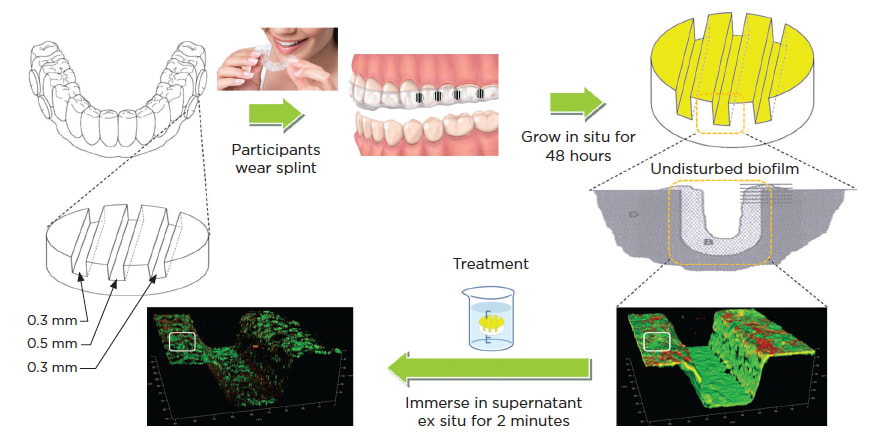
Figure 1
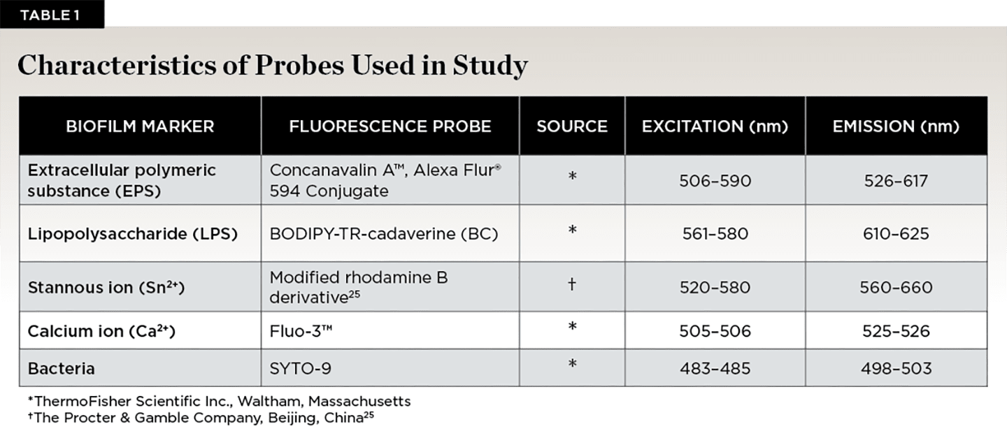
Table 1

Figure 2

Figure 3
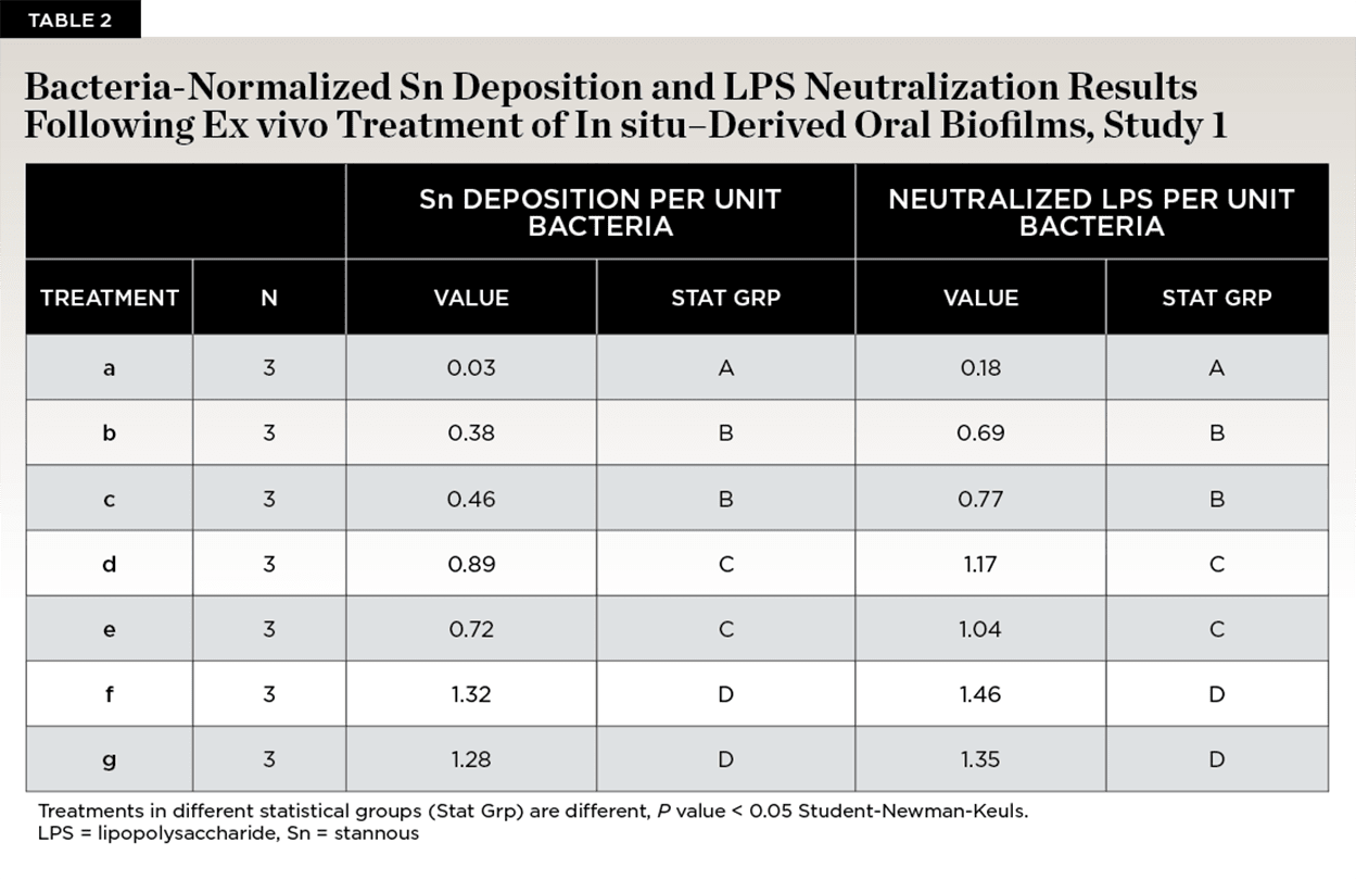
Table 2

Figure 4
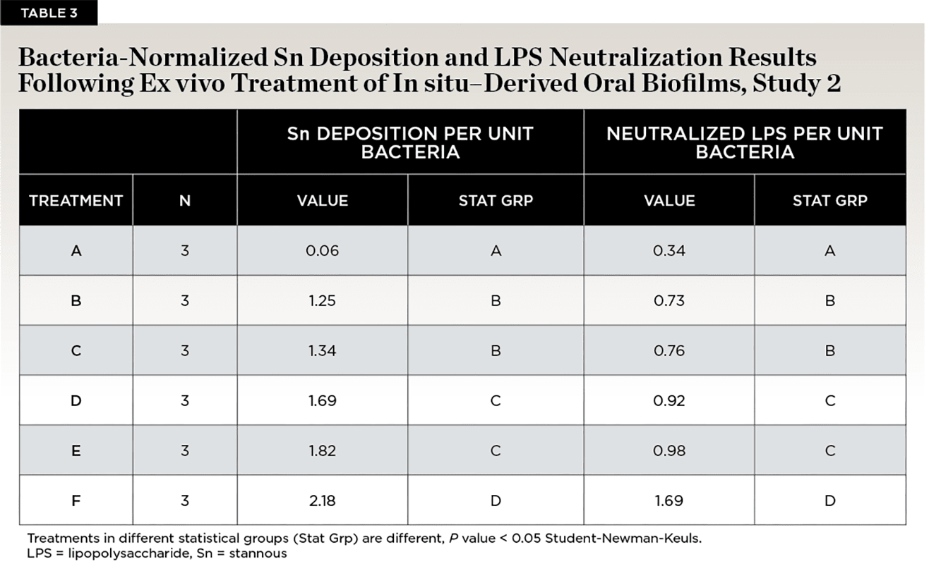
Table 3
