Abstract: This clinical case report describes a conservative technique that combines direct and indirect restorations to rehabilitate both maxillary central incisors following interdisciplinary orthodontic treatment. Demonstrating the integration of contemporary adhesive materials with digital workflows, the case illustrates the use of a direct composite restoration to correct the form and proportion of tooth No. 8 and a porcelain-fused-to-zirconia crown to restore tooth No. 9. A step-by-step clinical workflow is presented to help clinicians manage complex esthetic challenges while preserving natural tooth structure.
The maxillary central incisors represent a highly demanding area for esthetic rehabilitation. Color discrepancies, unfavorable tooth proportions, altered incisal edge position, and soft-tissue deficiencies can significantly affect a patient’s confidence while smiling and speaking. Establishing ideal incisal edge position, correcting tooth angulation, and creating proper interproximal contacts are essential components of successful anterior esthetics.
When carefully planned, interdisciplinary dentistry can improve both function and esthetic appearance while reducing treatment invasiveness. Advances in adhesive dentistry have expanded the role of both direct and indirect restorations as predictable options for restoring maxillary central incisors.
The authors prefer direct composite restorations for small to moderate defects where preservation of adjacent tooth structure is possible. Indirect restorations are considered when larger defects involve the entire facial surface or when replacing existing crowns. The following case report describes the combined use of direct and indirect restorative approaches following interdisciplinary orthodontic treatment to address varying deficiencies in the esthetic zone.
Case Presentation
A 30-year-old male patient presented for restorative consultation following completion of interdisciplinary orthodontic treatment that included maxillary rapid palatal expansion (MARPE) to address airway concerns, a constricted maxillary arch, and an unfavorable occlusion prior to restorative rehabilitation (Figure 1).
A digital smile design (DSD) analysis was performed to evaluate incisal edge position, the dental midline, and tooth proportions. A digital wax-up of teeth Nos. 8 and 9 was created to guide the correction of the existing diastema between the central incisors. The wax-up provided a reference for the mesio-incisal direct composite restoration on tooth No. 8 and served as a guide for provisionalization of tooth No. 9 following crown removal (Figure 2).
After 3D printing the diagnostic model, a palatal silicone putty matrix was fabricated to transfer the digital design into the clinical environment (Figure 3). Following etching and application of a universal dental adhesive, a two-layer composite technique using dentin and enamel A1 shades was performed. The tooth No. 8 direct restoration was finished and polished to achieve natural integration with the surrounding enamel.
Once tooth No. 8 was completed, the existing crown on tooth No. 9 was removed. The preparation was refined by eliminating sharp line angles and creating a smooth chamfer finish line extending approximately 1 mm subgingivally on the facial and proximal surfaces while remaining equigingival on the palatal surface. This preparation design facilitated an ideal emergence profile for provisional and definitive restorations while minimizing visibility of the restoration margin (Figure 4).
A size 000 retraction cord was placed, followed by digital impression acquisition. An intraoral photograph was taken with the selected A1 shade tab positioned adjacent to the prepared tooth, allowing the dental technician to evaluate both the abutment shade and final restoration requirements in the same image (Figure 5).
For a single maxillary central incisor requiring replacement of an existing crown, porcelain-fused-to-zirconia (PFZ) represents a reliable restorative option because it combines strength, masking ability, and customized facial characterization through layered porcelain (Figure 6).
Three weeks after the definitive impression, the PFZ crown was evaluated during the try-in appointment. The prepared tooth was cleaned using an intraoral air-abrasion unit with aluminum oxide particles and water before evaluation. A try-in paste was used to stabilize the crown while photographs were obtained to evaluate contour, texture, and shade integration (Figure 7). The images were reviewed with the patient prior to final cementation.
The crown was adhesively bonded using a dual-cure universal resin cement. A 3-second tack-cure technique was used to facilitate removal of excess cement before final polymerization (Figure 8).
A postoperative radiograph confirmed complete removal of residual cement. Static and dynamic occlusion were evaluated using thin articulating paper, and a final intraoral scan was obtained for fabrication of a 3D-printed occlusal guard.
Rationale for Treatment
Direct composite restoration remains the most conservative approach when defects can be restored while preserving healthy tooth structure. The primary challenge is creating an imperceptible transition between restorative material and natural enamel.
A dentin-and-enamel layering technique that respects the natural thickness and optical properties of tooth structure allows predictable esthetic integration. Shade selection should be completed at the beginning of the appointment before dehydration alters the tooth color. Clinical photography and spectrophotometers provide valuable tools for shade communication and restorative predictability.
Appropriate finishing burs and polishing systems are essential to reproduce enamel surface texture and gloss. Understanding the correct sequence and operating parameters allows direct composite restorations to achieve a surface appearance comparable to natural enamel.
For replacement of existing crowns in the esthetic zone, PFZ restorations provide a combination of strength, masking ability, and facial customization. Precise tooth preparation, careful soft-tissue management, and accurate digital impressions help improve communication with the laboratory and increase predictability.
A diagnostic and treatment-planning session with the laboratory team is an important step when managing complex esthetic cases. Whether treatment involves direct or indirect restorations, a facially driven digital wax-up should be considered a fundamental component of planning. It guides composite layering, preparation design, provisional restoration fabrication, and communication among the clinician, technician, and patient.
The integration of digital workflows and in-office 3D printing helps improve efficiency by allowing rapid fabrication of diagnostic models and silicone matrices. These matrices remain essential because they translate the digital treatment plan into a predictable clinical result.
Primary Products
Several key products (Kuraray, kuraraydental.com) were used in this case that were instrumental to achieving the successful outcome:
CLEARFIL™ Universal Quick Bond 2—This universal dental adhesive is designed to provide predictable bonding to enamel and dentin while simplifying adhesive procedures for both direct and indirect restorations.
CLEARFIL™ Majesty—A highly esthetic nanohybrid composite resin, Majesty offers excellent polishability, shade integration, and mechanical properties, making it suitable for anterior restorations using layered composite techniques.
Panavia™ SA Cement Universal—Designed for definitive cementation of indirect restorations, this dual-cure universal resin cement provides reliable adhesion to multiple restorative substrates while maintaining an efficient clinical workflow.
Conclusion
Successful rehabilitation of the maxillary central incisors requires careful integration of esthetic planning, restorative material selection, and preservation of healthy tooth structure. This case demonstrates how direct and indirect restorative approaches can be combined following interdisciplinary orthodontic treatment to address differing clinical needs within the same esthetic zone. A direct composite restoration provided a conservative solution for correcting the form and proportion of tooth No. 8, while a PFZ crown offered the strength, masking ability, and esthetic customization required for tooth No. 9. Digital smile design, a facially driven wax-up, 3D-printed models, and silicone matrices facilitated transfer of the treatment plan to the clinical setting and enhanced communication among the clinician, laboratory technician, and patient. By selecting the least invasive appropriate treatment for each tooth and integrating contemporary adhesive materials with digital workflows, clinicians can achieve predictable esthetic outcomes while maximizing preservation of the patient’s natural dentition.
DISCLOSURE
This article was commercially supported by Kuraray.
ABOUT THE AUTHORS
Julian Conejo, DMD
Private Practice, Newtown Square and Glen Mills, Pennsylvania
Marianna Evans, DMD
Private Practice, Newtown Square and Glen Mills, Pennsylvania
David Anderson, CDT
Master Ceramist, Lotus Collection by Hybridge Lab, Rochester, New York
References
1. Conejo J. Current adhesive protocols for indirect ceramic restorations. Compend Contin Educ Dent. 2022;43(9):592-594.
2. Blatz MB, Alammar A, Rojas F, Conejo J. How to bond to current CAD/CAM ceramics. Compend Contin Educ Dent. 2023;44(10):560-565.
3. Eggmann F, Ayub JM, Conejo J, Blatz MB. Deep margin elevation—present status and future directions. J Esthet Restor Dent. 2023;35(1):26-47.
4. Eggmann F, Mante FK, Ayub JM, et al. Influence of universal adhesives and silane coupling primer on bonding performance to CAD/CAM resin-based composites: a laboratory investigation. J Esthet Restor Dent. 2024;36(4):620-631.
5. Almalki A, Conejo J, Wünsche A, et al. Digital smile design and fabrication of CAD/CAM restorations in a complex esthetic case. Compend Contin Educ Dent. 2022;43(10):664-668.
6. Blatz MB, Vonderheide M, Conejo J. The effect of resin bonding on long-term success of high-strength ceramics. J Dent Res. 2018;97(2):132-139.
7. Evans M. Airway and periodontal rationale for orthopedic jaw development in the primary and early mixed dentition. Continuing Dental Education course at University of Pennsylvania School of Dental Medicine.
Figures and Images
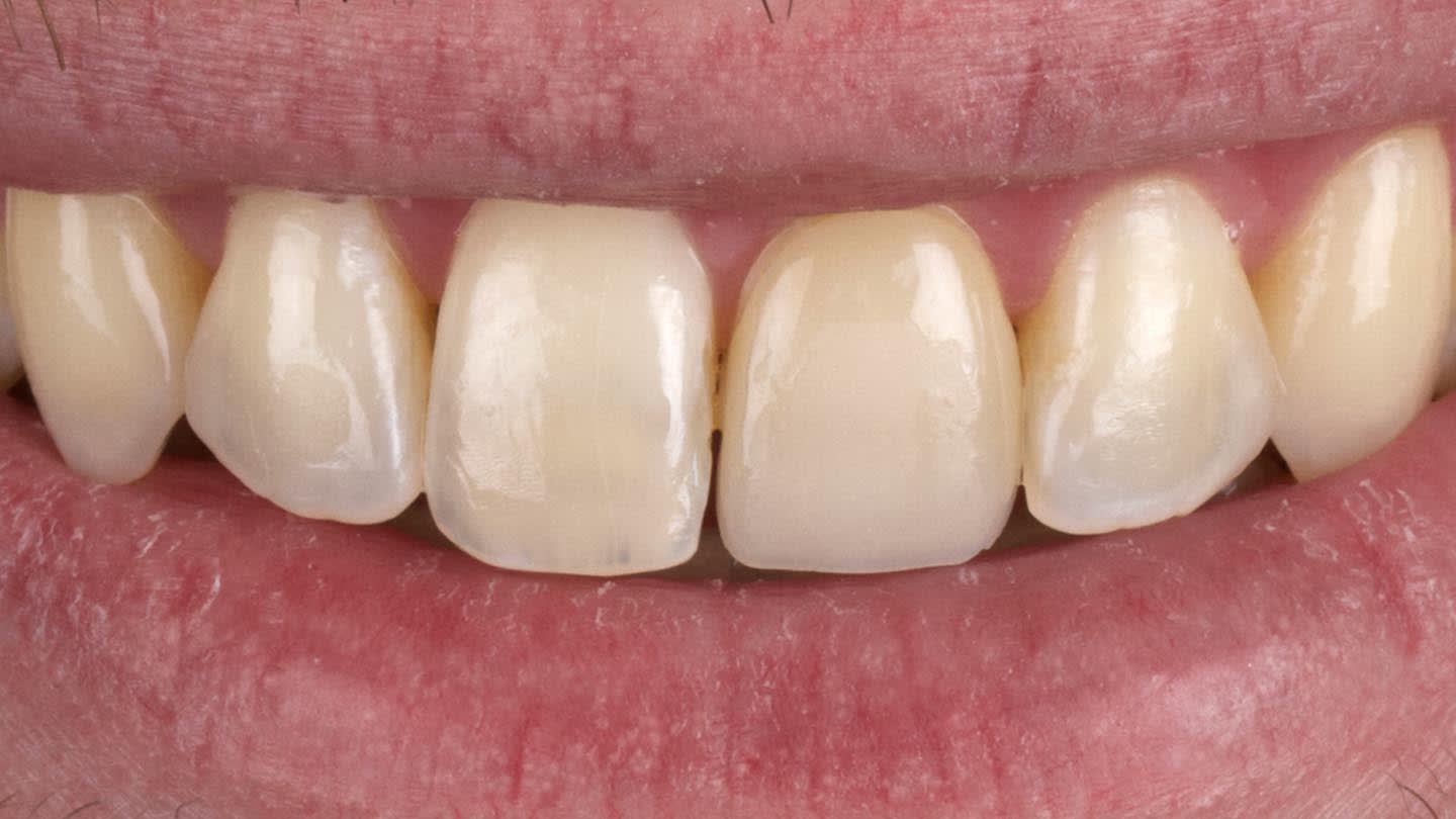
Figure 1
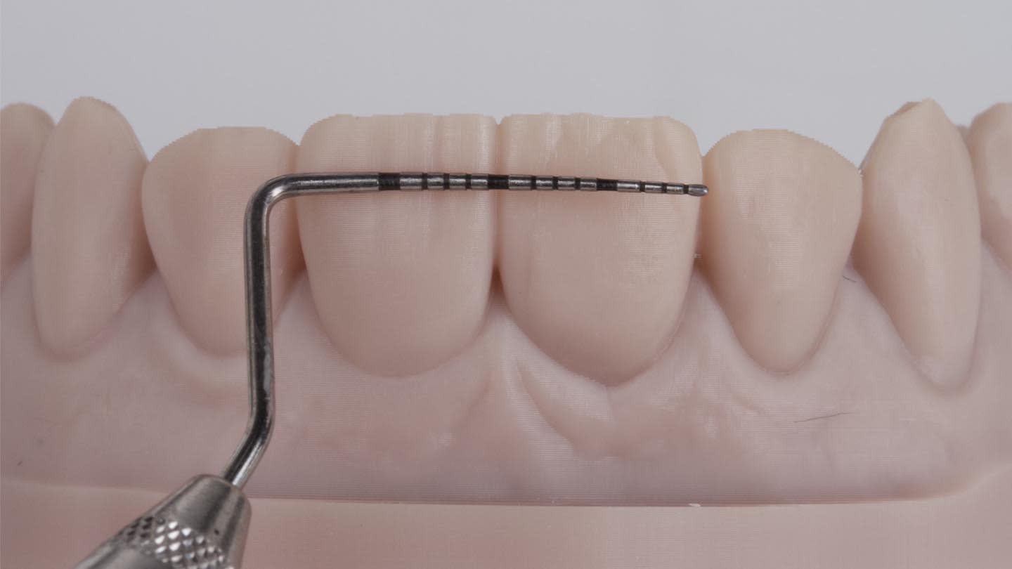
Figure 2
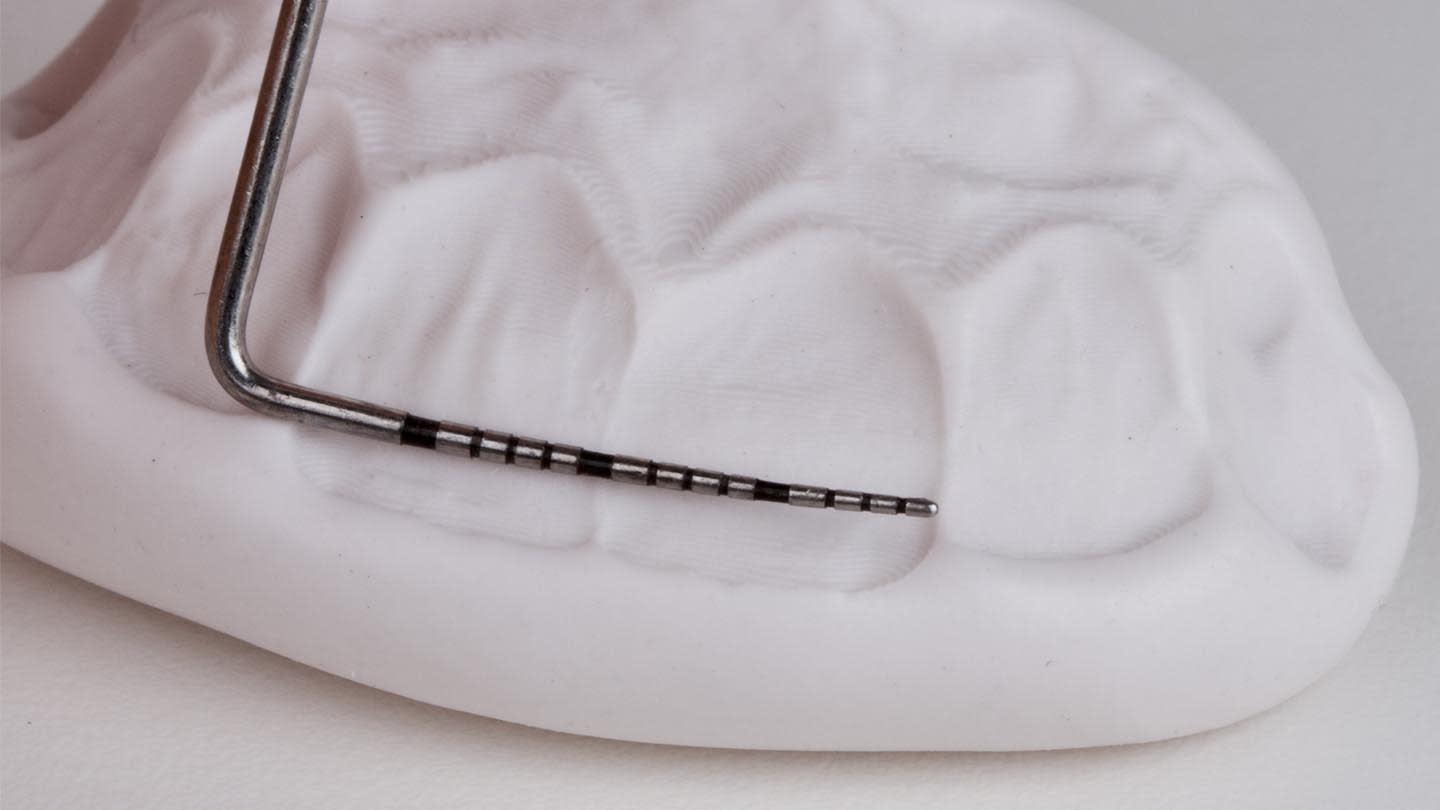
Figure 3
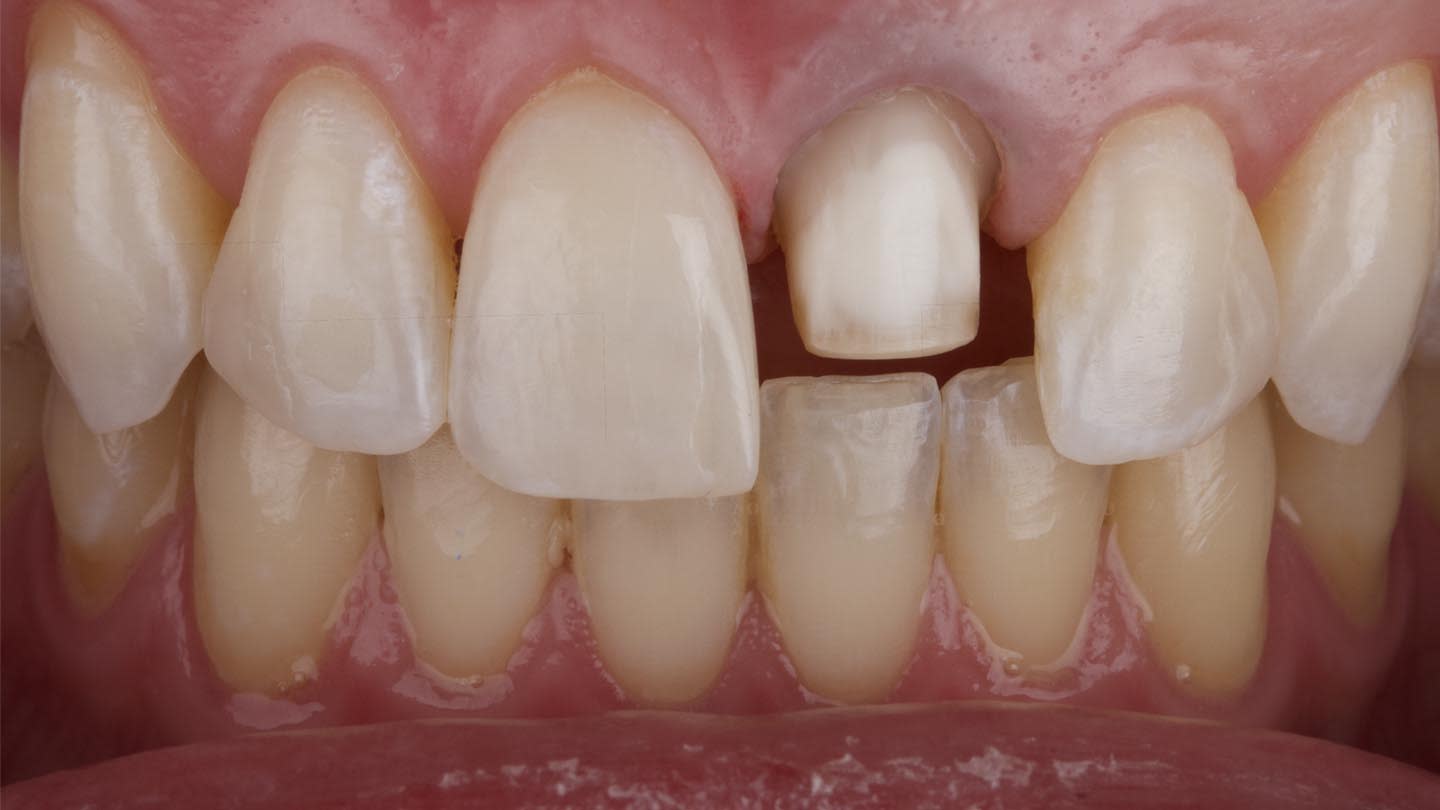
Figure 4
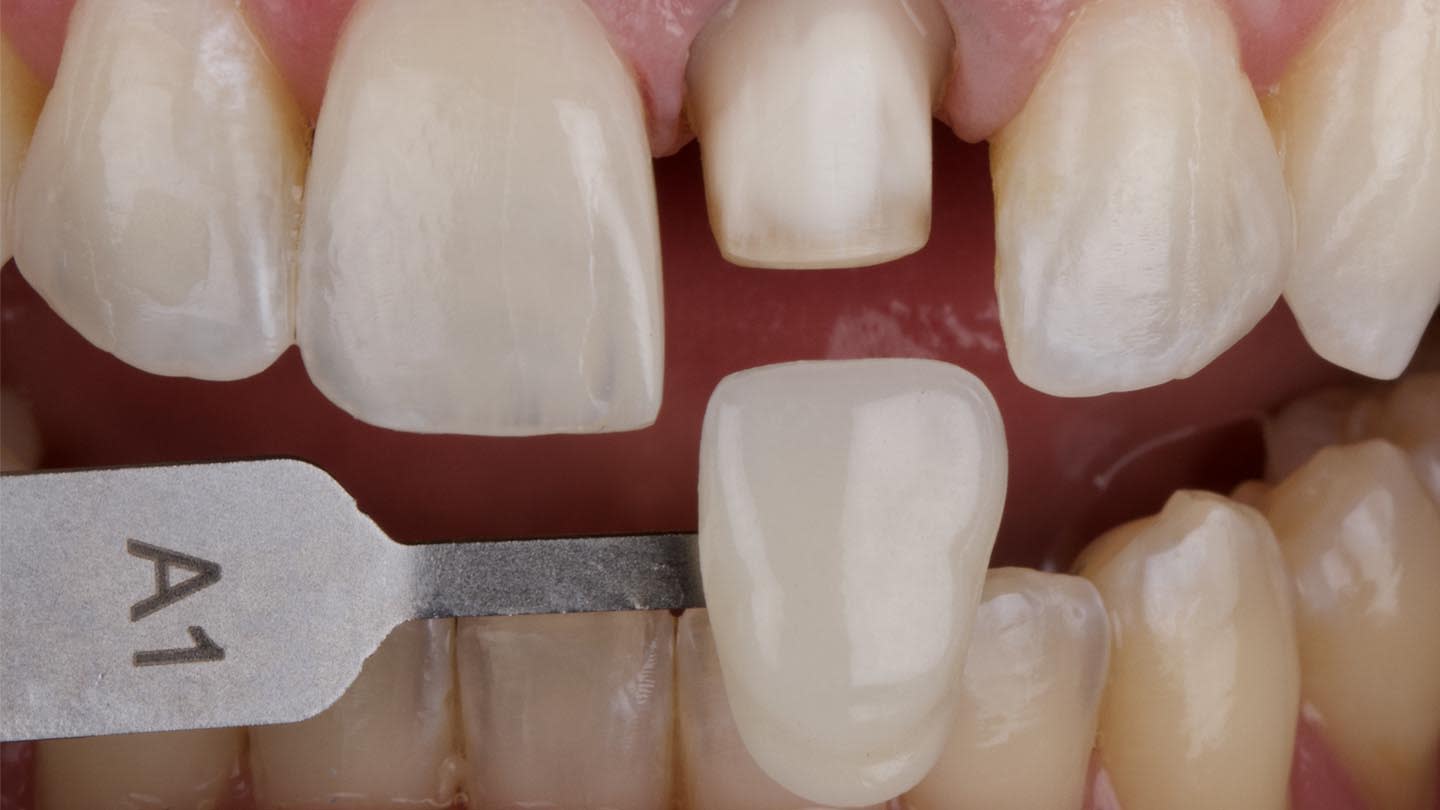
Figure 5
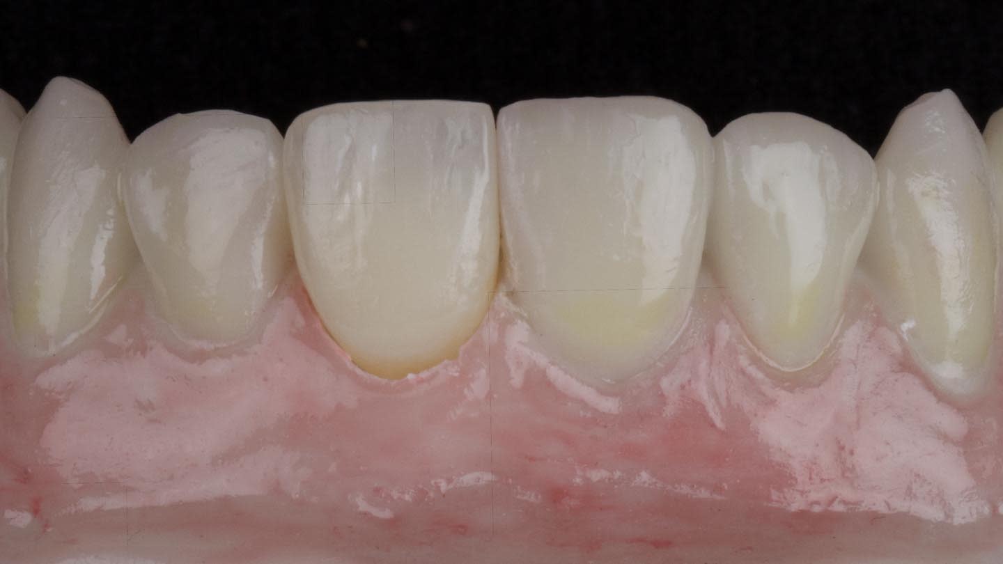
Figure 6
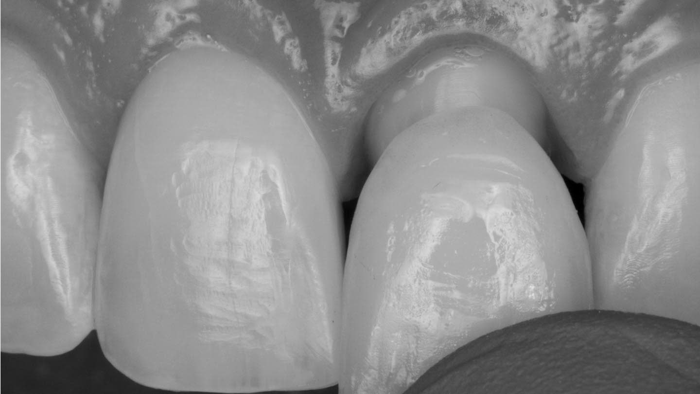
Figure 7
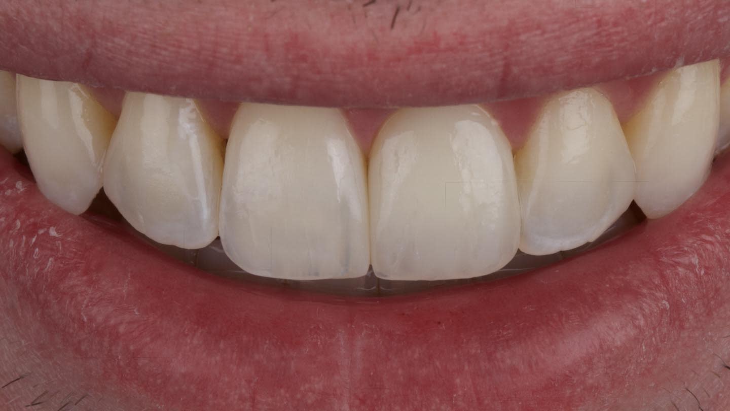
Figure 8