Abstract: Contemporary pulpotomy in primary molars reflects a shift from the use of devitalizing agents toward biologically driven vital pulp therapy. Advances in pulpal biology and biomaterials, particularly calcium silicate–based medicaments, support healing and preservation of radicular pulp vitality, leading to improved clinical outcomes. Current recommendations emphasize accurate diagnosis through integration of patient history, clinical findings, and radiographic evaluation, along with careful case selection. Achievement of a durable coronal seal remains essential and is most predictably attained with full-coverage restorations. As described in this case report, when modern bioactive materials are combined with appropriate restorative protocols, pulpotomy is a reliable and effective treatment for primary teeth with deep carious lesions.
For many clinicians, pulpotomy in primary molars has long been a familiar and dependable procedure. The typically reliable treatment utilizes well-known materials, and outcomes are generally acceptable. However, advances in dental biomaterials and an expanding body of clinical evidence have prompted a reassessment of traditional approaches to pulp therapy in children. Contemporary pediatric dentistry increasingly emphasizes biologically driven treatment strategies that prioritize preservation of pulp vitality and support the natural healing capacity of dental tissues.1,2
This shift reflects a deeper understanding of pulpal biology, inflammation, and the importance of an effective coronal seal in determining treatment success. Updated recommendations from the American Academy of Pediatric Dentistry for vital pulp therapy outline diagnostic decision pathways and provide evidence for the most successful forms of treatment, including indirect pulp therapy, direct pulp therapy, and pulpotomy. These guidelines also address material selection for managing primary teeth with deep caries when the radicular pulp remains vital.1 Rather than relying on materials that primarily devitalize tissue, current protocols encourage the use of bioactive medicaments capable of supporting repair and maintaining pulpal health.

S Series Implant Portfolio
The progression of pulpotomy medicaments illustrates how this evolution has unfolded. Calcium hydroxide, introduced in the early twentieth century, was initially embraced because of its antimicrobial properties and high pH. While biologically appealing in theory, clinical experience in primary teeth revealed inconsistent outcomes and a relatively high incidence of internal root resorption.3 Formocresol subsequently became widely adopted and served as the benchmark pulpotomy medicament for decades due to its predictable clinical performance and ease of use. Despite these advantages, formocresol functions by devitalizing pulp tissue rather than supporting regeneration, and concerns regarding formaldehyde exposure have prompted clinicians to consider alternative materials.4
Ferric sulfate was later introduced as a hemostatic agent that avoided formaldehyde exposure. Although straightforward to apply and initially promising, longer-term studies involving ferric sulfate demonstrated variable success rates as well as internal resorption and radiographic or clinical failure in some cases.5 These limitations reinforced the need for materials capable of supporting biologic healing rather than simply controlling hemorrhage or devitalizing tissue.
The introduction of mineral trioxide aggregate (MTA) in the 1990s represented a pivotal development in vital pulp therapy. MTA and other calcium silicate–based biomaterials create a biologically favorable environment through sustained alkalinity, calcium ion release, and stimulation of hard-tissue formation. Numerous clinical trials and systematic reviews have demonstrated high success rates when these materials are used in pulpotomy procedures.2,6 More recently, premixed calcium silicate formulations, such as EdgeUtopia™ Root Repair Material (EdgeEndo, edgeendo.com) (Figure 1), have been introduced to retain the biologic advantages of earlier bioceramics while improving handling characteristics and clinical efficiency.
Along with advances in pulpotomy medicaments, restorative strategies have also evolved. Full-coverage restorations, most commonly stainless steel crowns and more recently esthetic zirconia pediatric crowns, remain the recommended definitive restoration for pulpotomized primary molars because of their durability and ability to maintain a longterm coronal seal.1 When biologically compatible materials and appropriate restorative protocols are combined, pulpotomy in the primary dentition becomes a predictable procedure that supports both tooth retention and pulpal health.
Rationale and Diagnosis
The objective of pulpotomy in primary teeth is to remove inflamed coronal pulp tissue while preserving the vitality and function of the remaining radicular pulp. When successful, this procedure enables the tooth to remain in the arch until natural exfoliation, helping to maintain arch length, masticatory function, esthetics, and appropriate eruption guidance for the developing permanent successor.7
Appropriate case selection begins with a careful diagnosis that integrates patient history, clinical examination, and radiographic findings. In pediatric dentistry, the pain history is often provided by a parent or caregiver and may not always be precise. Even when children describe their own symptoms, they may be unreliable historians or may have adapted to chronic discomfort without recognizing it as abnormal. Targeted questions can help, but they must always be interpreted alongside clinical and radiographic findings.
Clinicians should ask when pain occurs, whether during the day or at night. Nocturnal pain is often associated with more advanced pulpal inflammation and may reduce the likelihood of pulpotomy success. Spontaneous pain, particularly pain that occurs at night, is commonly associated with irreversible pulpitis and may not respond well to vital pulp therapy.8 The duration of symptoms is also relevant. Transient pain that resolves quickly is generally more favorable, whereas prolonged lingering pain may indicate deeper pulpal involvement. Finally, clinicians should ask how the pain responds to analgesics. Relief with analgesics may suggest a less advanced inflammatory state, whereas lack of relief may indicate a more progressed pulpitis that may not respond favorably to vital pulp therapy.
Radiographic evaluation is equally important. Particular attention should be directed to the furcation region when examining primary molars, as the pulp chamber of primary molars communicates with the furcation through accessory canals, making this the most common site for pathologic radiolucency. The presence of furcation radiolucency, pathologic root resorption, or widening of the periodontal ligament space may indicate pulpal necrosis and contraindicate pulpotomy. However, the absence of radiographic findings does not guarantee pulpal health, because radiographic changes often develop later in the disease process.9
Clinical findings also guide diagnosis. Indications for pulpotomy typically include deep caries approaching the pulp or a carious pulp exposure with controlled hemorrhage and no signs of irreversible pulpitis. Contraindications include pathologic mobility or a tooth that cannot be predictably restored. Restorability is therefore a critical consideration. Even when pulpal conditions appear favorable, pulpotomy should not be undertaken if the tooth cannot be sealed effectively. Time to exfoliation must also be considered. Primary teeth are transitional, and when the remaining lifespan of the tooth is short, extraction rather than vital pulp therapy may be the more appropriate option.
Restoration Considerations
Restoration following vital pulp therapy, specifically pulpotomy, is a critical component of treatment success. Stainless steel crowns have historically been considered the gold standard restoration because they provide full coronal coverage and reduce the risk of microleakage. Numerous studies have demonstrated higher survival rates when pulpotomized primary molars are restored with full-coverage restorations rather than intracoronal restorations.10,11
Advances in restorative materials and adhesive technologies continue to improve the ability to achieve durable coronal seals. Regardless of the material used, the quality of the final restoration remains a central factor in the long-term success of pulpotomy therapy.
Case Study
A 2-year-and-8-month-old boy presented with severe early childhood caries affecting all eight primary molars and the maxillary incisors. The parent reported no complaints of pain but described the child’s teeth as being “in pretty bad shape.” Clinical examination revealed soft tissues within normal limits. Radiographic examination was also within normal limits with no evidence of furcation pathology. A deep occlusal carious lesion approximating the pulp was noted on a maxillary first primary molar, which was selected for pulpotomy treatment (Figure 2). The anterior teeth were determined to be nonrestorable because of the extent of structural destruction.
Due to the patient’s young age and the severity and distribution of decay, comprehensive treatment would be completed in the operating room under general anesthesia. This entailed full oral rehabilitation, including full-coverage pediatric preformed crowns, intracoronal restorations, and extractions.
After induction of general anesthesia, the maxillary first primary molar selected for pulpotomy treatment was isolated with a rubber dam (Latex Black Dental Dam, Sanctuary, sanctuary-dental.com) to ensure optimal moisture control and procedural safety. The rubber dam was placed using a slot cut technique, and a W2A clamp (Ivory®, Kulzer, kulzerus.com) was secured to the second primary molar to provide stable isolation (Figure 3).
Initial tooth preparation was performed in anticipation of placement of a stainless steel crown. Occlusal reduction of approximately 1 mm was completed using a high-speed handpiece (NSK, nsk-dental.com) and a NeoDiamond 1116.6C bur (Microcopy, microcopydental.com). The occlusal preparation is shown in Figure 4.
Access to the pulp chamber was then created, and the roof of the chamber was removed (Figure 5). Caries excavation and coronal pulp amputation were performed using a slow-speed handpiece (NSK) with a large #8 round RA bur (Microcopy). Coronal pulp removal is demonstrated in Figure 6.
Hemostasis was achieved using sterile saline irrigation (Figure 7) followed by application of gentle pressure with a moistened cotton pellet (Figure 8). Controlled bleeding from the radicular pulp tissue was observed (Figure 9), indicating a vital radicular pulp suitable for pulpotomy therapy.
A bioactive calcium silicate pulpotomy medicament (EdgeUtopia Root Repair Material) was placed directly over the canal orifices (Figure 10). The material was adapted using a nonstick instrument (LM Dark Diamond™, LM-Dental, lm-dental.com) (Figure 11) and positioned to fill the coronal pulp chamber (Figure 12).
Following medicament placement, a stainless steel crown (Solventum, solventum.com) was cemented using a glass ionomer luting cement (GC FujiCEM® Evolve, GC America, gc.dental/america). The completed restoration is shown in Figure 13.
Conclusion
Contemporary pulpotomy in the primary dentition reflects a shift toward biologically driven treatment strategies that prioritize preservation of pulp vitality. Advances in calcium silicate–based materials have expanded clinicians’ ability to perform vital pulp therapy in a manner that supports natural healing while maintaining procedural efficiency. Successful outcomes, however, depend on more than the medicament alone. Accurate diagnosis remains fundamental and requires thoughtful interpretation of patient history, clinical findings, and radiographic assessment. Equally critical is the achievement of a durable coronal seal, most predictably accomplished with full-coverage restorations such as stainless steel crowns.
When appropriate case selection, modern bioactive materials, and definitive restorative protocols are combined, pulpotomy becomes a reliable and efficient treatment option for primary molars with deep carious lesions. As materials and techniques continue to evolve, clinicians have the opportunity to deliver care that is both predictable and aligned with biologic principles that support pulpal health and tooth preservation.
DISCLOSURE
This article was commercially supported by EdgeEndo.
ABOUT THE AUTHOR
Carla Cohn, DMD
Continuing Dental Educator, Part-time Clinical Instructor Pediatric Dentistry, Gerald Niznick College of Dentistry, University of Manitoba; Private Practice, Winnipeg, Manitoba, Canada
References
1. Coll JA, Dhar V, Chen CY, et al. Use of vital pulp therapies in primary teeth 2024. Pediatr Dent. 2024;46(1):13-26.
2. Coll JA, Seale NS, Vargas K, et al. Primary tooth vital pulp therapy: a systematic review and meta-analysis. Pediatr Dent. 2017;39(1):16-123.
3. Fuks AB. Current concepts in vital primary pulp therapy. Eur J Paediatr Dent. 2002;3(3):115-120.
4. Milnes AR. Is formocresol obsolete? A fresh look at the evidence concerning safety issues. J Endod. 2008;34(7 suppl):S40-S46.
5. Peng L, Ye L, Guo X, et al. Evaluation of formocresol versus ferric sulfate primary molar pulpotomy: a systematic review and meta-analysis. Int Endod J. 2007;40(10):751-757.
6. Holan G, Eidelman E, Fuks AB. Long term evaluation of pulpotomy in primary molars using mineral trioxide aggregate or formocresol. Pediatr Dent. 2005;27(2):129-136.
7. Fuks AB. Vital pulp therapy with new materials for primary teeth: new directions and treatment perspectives. Pediatr Dent. 2008;30(3):211-219.
8. American Academy of Pediatric Dentistry. Pulp therapy for primary and immature permanent teeth. The Reference Manual of Pediatric Dentistry. Chicago, IL: American Academy of Pediatric Dentistry; 2025:487-496.
9. Duggal MS, Nooh A, High A. Response of the primary pulp to inflammation: a review of the Leeds studies and challenges for the future. Eur J Paediatr Dent. 2002;3(3):111-114.
10. Seale NS, Randall R. The use of stainless steel crowns: a systematic literature review. Pediatr Dent. 2015;37(2):145-160.
11. Innes NPT, Ricketts D, Chong LY, et al. Preformed crowns for decayed primary molar teeth. Cochrane Database Syst Rev. 2015;2015(12):CD005512.
Figures and Images

Figure 1
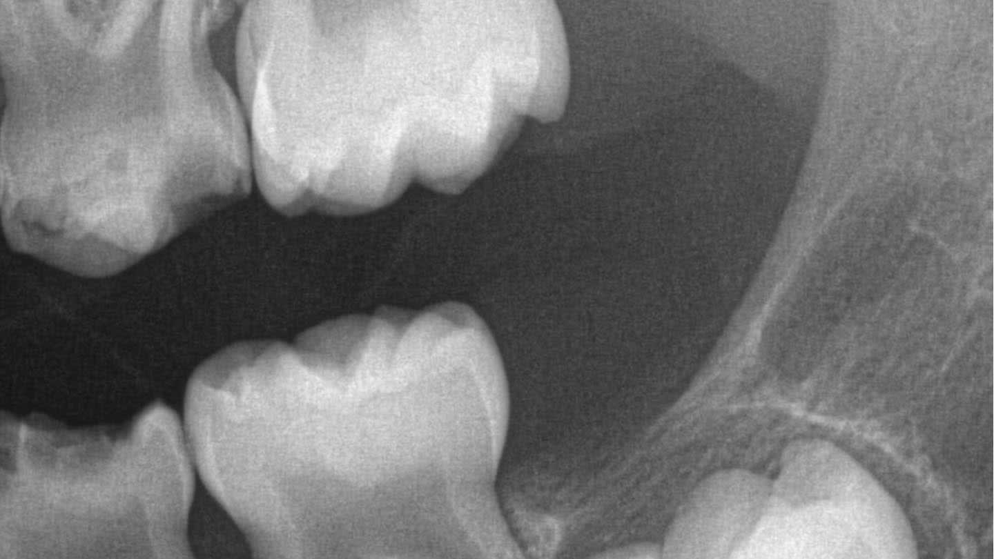
Figure 2

Figure 3

Figure 4
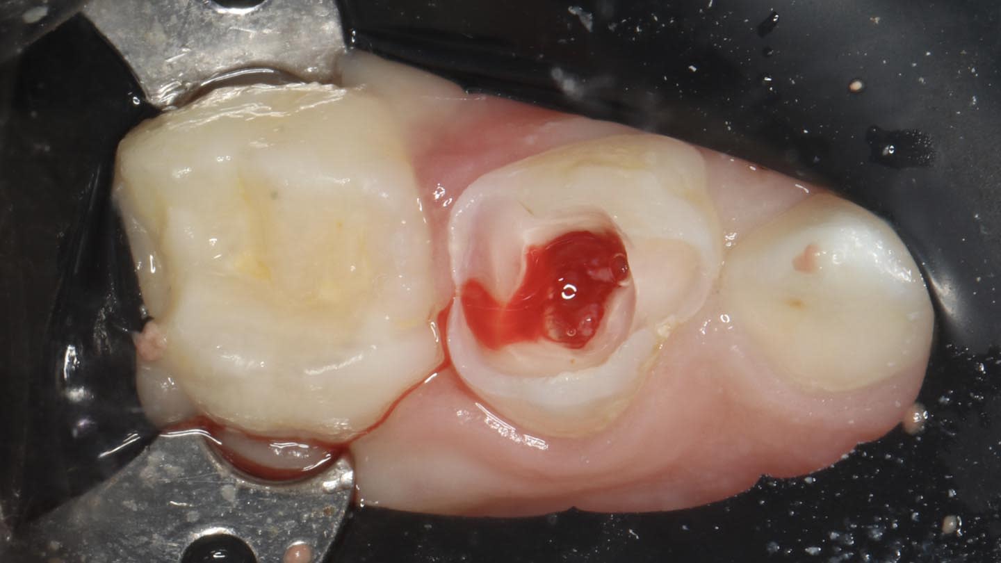
Figure 5
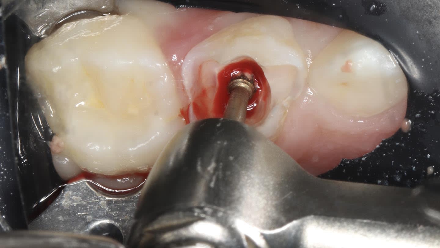
Figure 6

Figure 7
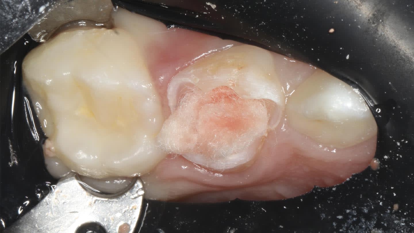
Figure 8
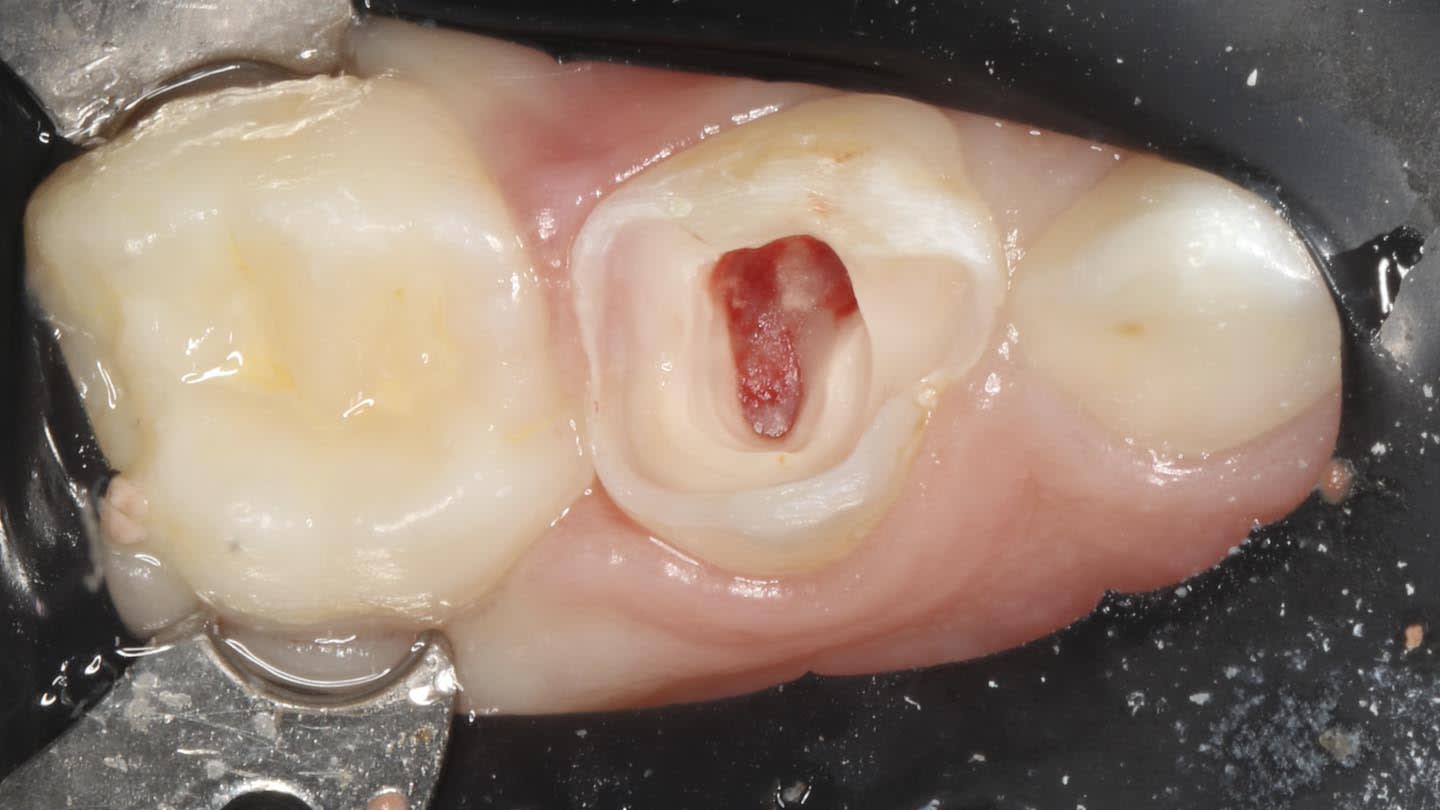
Figure 9

Figure 10
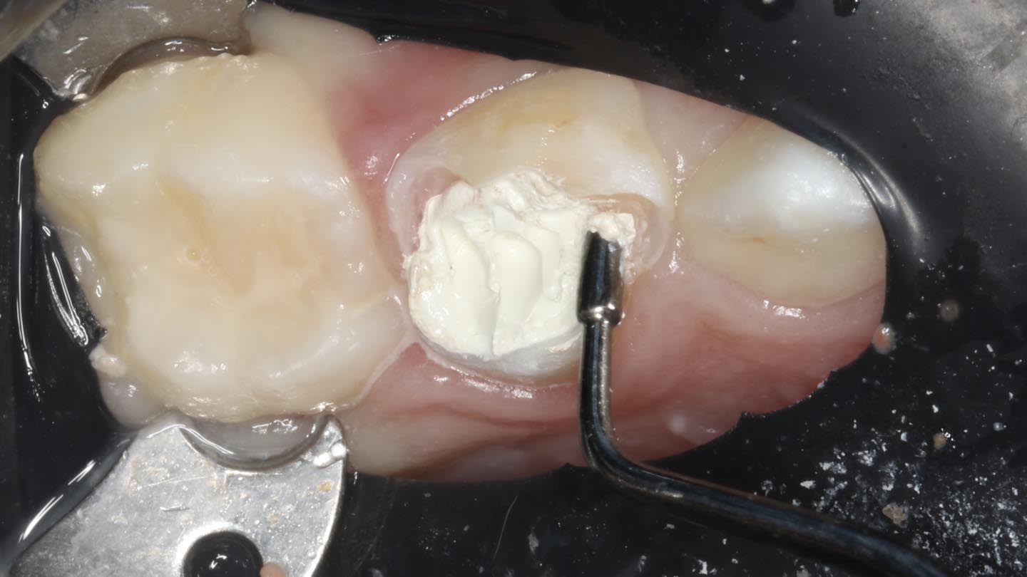
Figure 11

Figure 12

Figure 13
