In this case, the adolescent patient presented with peg lateral incisors, and the maxillary right canine and first premolar were in swapped positions. The orthodontist (Overlake Orthodontics) did an excellent job positioning the teeth to allow for post-orthodontic restorations with a modified technique using a single-shade composite system (OMNICHROMA, Tokuyama Dental,
omnichroma.com). The treatment goals were to make tooth No. 5, which was in the No. 6 position, look like a canine, and make the peg lateral incisors appear normal in size and shape. First, the patient was able to whiten the teeth (Zoom!, Philips), during which time a diagnostic wax-up and silicone putty index were fabricated in preparation for the bonding appointment. A carbon dioxide (CO2) laser was used without anesthetic to contour the gingival zeniths and prepare the surface of the enamel for bonding. The layering technique used for this single-shade composite system mimics that of a polychromatic layering composite: a palatal shell with OMNICHROMA, followed by OMNICHROMA BLOCKER to replicate dentin, another thin layer of OMNICHROMA, colored resin stains (Estelite® Color, Tokuyama Dental), and a final topcoat of OMNICHROMA. As seen in this case presentation, following this technique with a single-shade composite system enabled highly esthetic results.
KEY TAKEAWAYS
Indicated for both direct anterior and posterior restorations, OMNICHROMA single-shade composite and OMNICHROMA BLOCKER can be used in a layering technique that mimics that of a polychromatic layering composite to achieve highly esthetic outcomes.
Utilizing Smart Chromatic Technology and supra-nano spherical fillers, OMNICHROMA universal composite is capable of shade-matching any tooth color from A1 to D4, streamlining the restorative process.
In addition to exceptional color matching, OMNICHROMA single-shade universal composite offers strength, durability, versatility, and high polishability, along with excellent physical-mechanical properties.
Kevin Brown, DDS
Private Practice, Bellevue, Washington; Accreditation, American Academy of Cosmetic Dentistry
Figures and Images
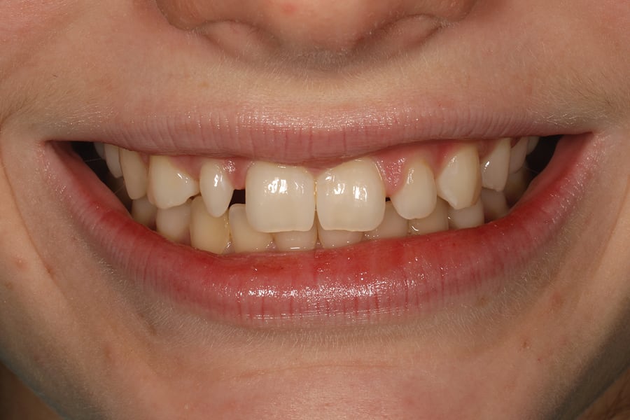
Figure 1
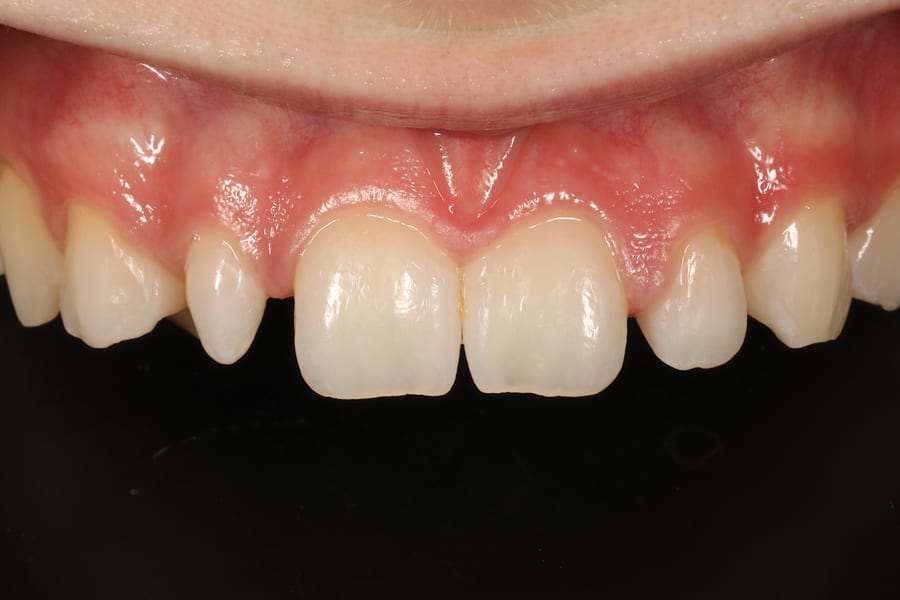
Figure 2
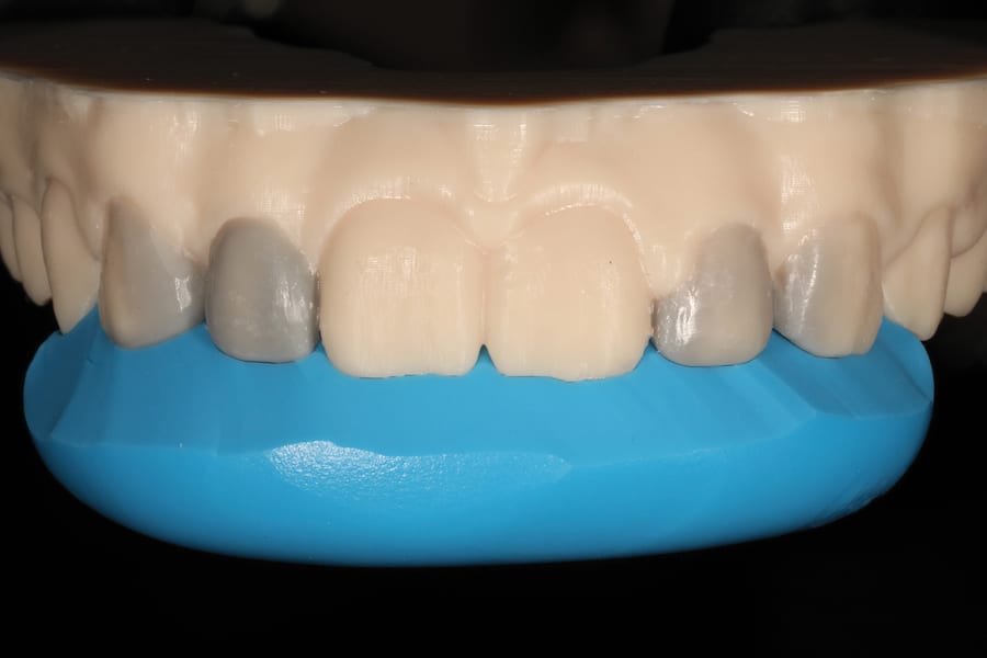
Figure 3
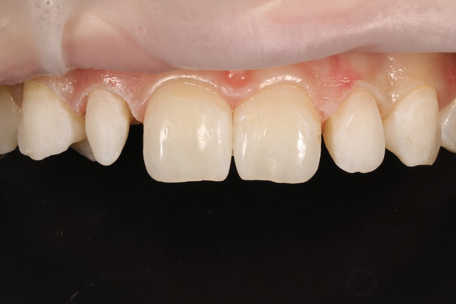
Figure 4
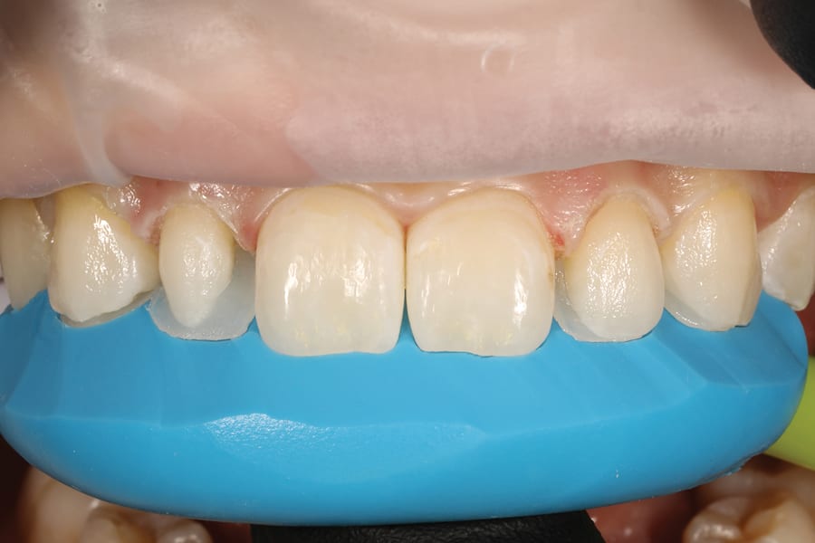
Figure 5
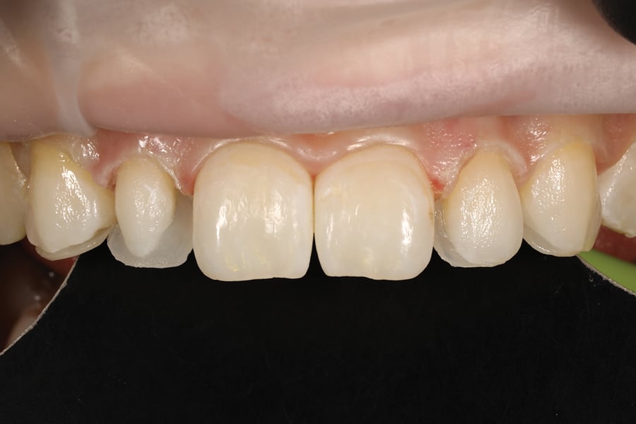
Figure 6
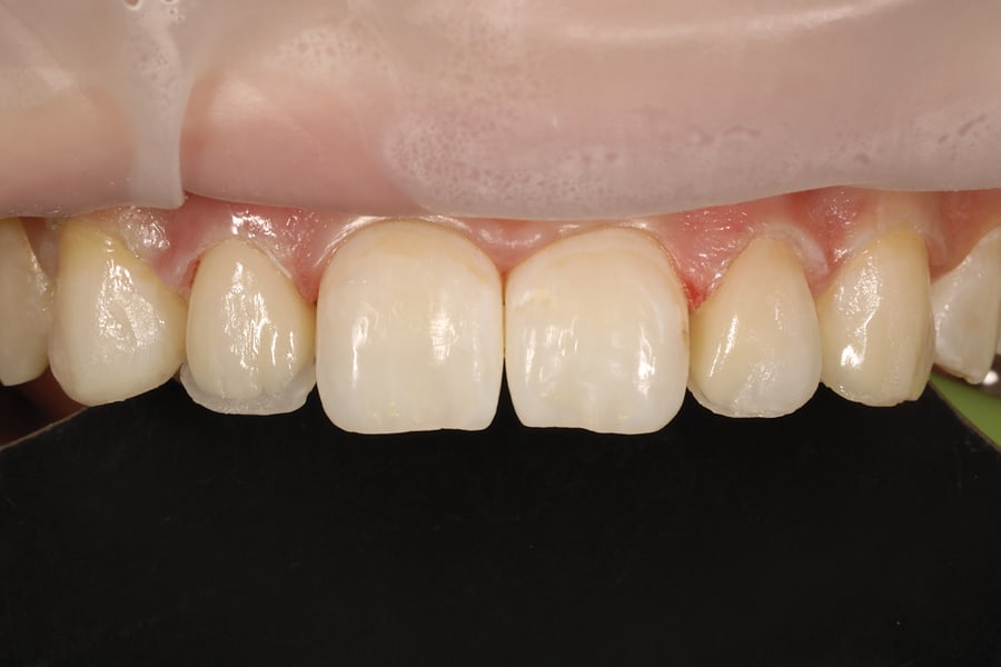
Figure 7
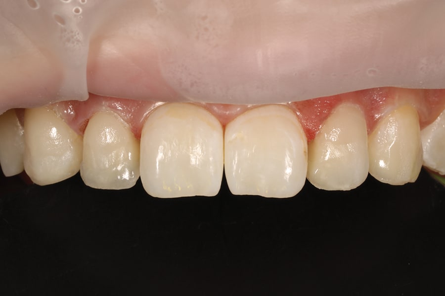
Figure 8
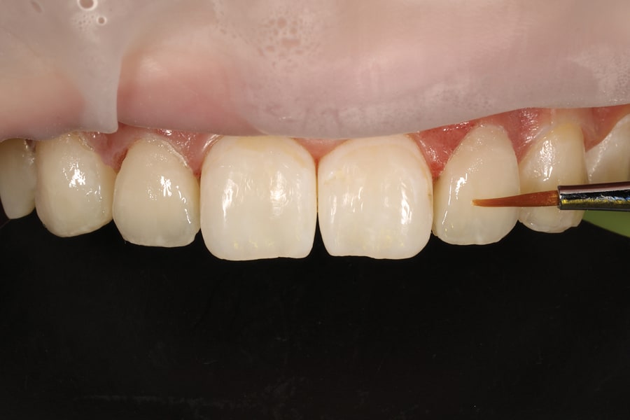
Figure 9
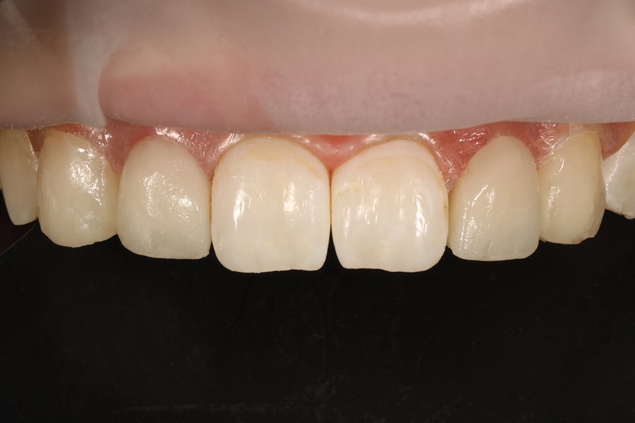
Figure 10
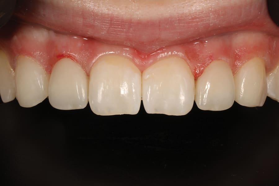
Figure 11
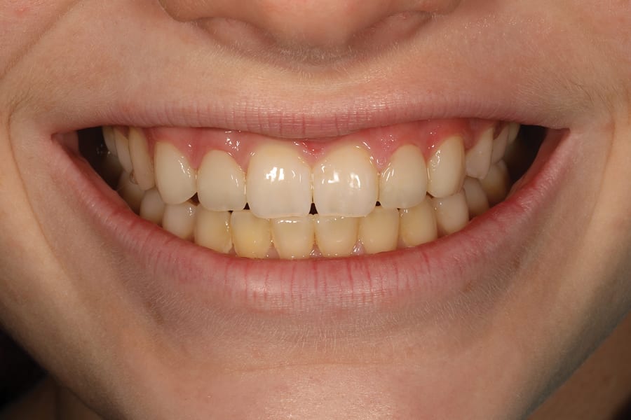
Figure 12
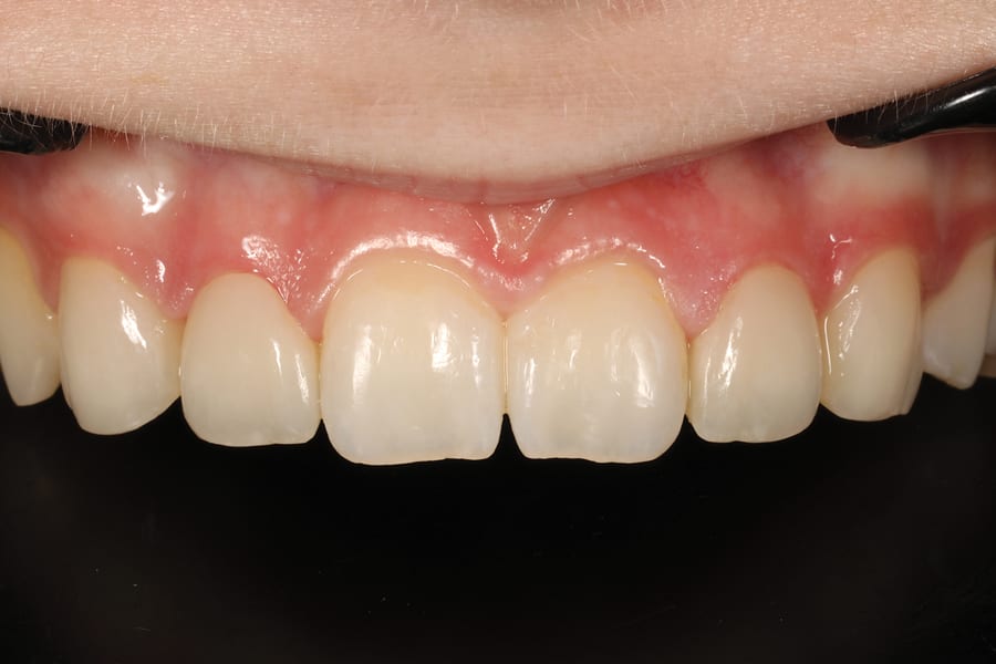
Figure 13
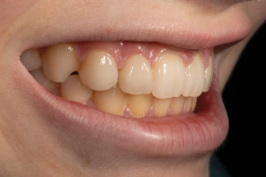
Figure 14